 Movie
Movie Controller

[English] 日本語
 Yorodumi
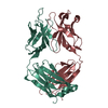
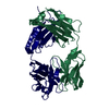
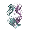
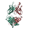
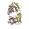
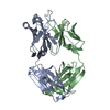
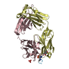
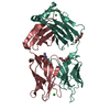
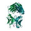
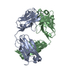
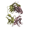
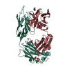
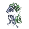
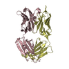
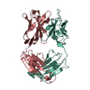
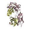
- PDB-1p7k: Crystal structure of an anti-ssDNA antigen-binding fragment (Fab)... -
+ Open data

- Basic information
| Entry | Database: PDB / ID: 1p7k | ||||||
|---|---|---|---|---|---|---|---|
| Title | Crystal structure of an anti-ssDNA antigen-binding fragment (Fab) bound to 4-(2-Hydroxyethyl)piperazine-1-ethanesulfonic acid (HEPES) | ||||||
 Components Components |
| ||||||
 Keywords Keywords |  IMMUNE SYSTEM / FAB / ANTIBODY / ANTI-DNA ANTIBODY / AUTOANTIBODY / LUPUS / HEPES IMMUNE SYSTEM / FAB / ANTIBODY / ANTI-DNA ANTIBODY / AUTOANTIBODY / LUPUS / HEPES | ||||||
| Function / homology |  Function and homology information Function and homology informationhumoral immune response mediated by circulating immunoglobulin / phagocytosis, recognition / positive regulation of type IIa hypersensitivity / positive regulation of type I hypersensitivity / antibody-dependent cellular cytotoxicity / Fc-gamma receptor I complex binding / immunoglobulin complex / phagocytosis, engulfment / IgG immunoglobulin complex / immunoglobulin mediated immune response ...humoral immune response mediated by circulating immunoglobulin / phagocytosis, recognition / positive regulation of type IIa hypersensitivity / positive regulation of type I hypersensitivity / antibody-dependent cellular cytotoxicity / Fc-gamma receptor I complex binding / immunoglobulin complex / phagocytosis, engulfment / IgG immunoglobulin complex / immunoglobulin mediated immune response / immunoglobulin complex, circulating / immunoglobulin receptor binding / positive regulation of phagocytosis / B cell differentiation / complement activation, classical pathway / antigen binding / positive regulation of immune response / antibacterial humoral response / adaptive immune response / defense response to bacterium / external side of plasma membrane / extracellular space / plasma membrane / cytoplasmSimilarity search - Function | ||||||
| Biological species |  Mus musculus (house mouse) Mus musculus (house mouse) | ||||||
| Method | X-RAY DIFFRACTION / SYNCHROTRON / MOLECULAR REPLACEMENT / Resolution: 1.75 Å | ||||||
 Authors Authors | Schuermann, J.P. / Henzl, M.T. / Deutscher, S.L. / Tanner, J.J. | ||||||
 Citation Citation | Journal: Proteins / Year: 2004 Title: Structure of an anti-DNA fab complexed with a non-DNA ligand provides insights into cross-reactivity and molecular mimicry. Authors: Schuermann, J.P. / Henzl, M.T. / Deutscher, S.L. / Tanner, J.J. #1: Journal: J.Mol.Biol. / Year: 2001Title: CRYSTAL STRUCTURE OF AN ANTIGEN-BINDING FRAGMENT BOUND TO SINGLE-STRANDED DNA Authors: TANNER, J.J. / KOMISSAROV, A.A. / DEUTSCHER, S.L. #2: Journal: Acta Crystallogr.,Sect.D / Year: 2000Title: CRYSTALLIZATION AND MOLECULAR REPLACEMENT STUDIES OF A RECOMBINANT ANTIGEN-BINDING FRAGMENT COMPLEXED WITH SINGLE-STRANDED DNA Authors: PREWITT, S.P. / KOMISSAROV, A.A. / DEUTSCHER, S.L. / TANNER, J.J. | ||||||
| History |
|
- Structure visualization
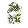
| Structure viewer | Molecule: MolmilJmol/JSmol |
|---|
- Downloads & links
-Download
| PDBx/mmCIF format | 1p7k.cif.gz | 203.2 KB | Display | PDBx/mmCIF format |
|---|---|---|---|---|
| PDB format | pdb1p7k.ent.gz | 159.1 KB | Display | PDB format |
| PDBx/mmJSON format | 1p7k.json.gz | Tree view | PDBx/mmJSON format | |
| Others |  Other downloads Other downloads |
-Validation report
| Arichive directory | https://data.pdbj.org/pub/pdb/validation_reports/p7/1p7kftp://data.pdbj.org/pub/pdb/validation_reports/p7/1p7k | HTTPS FTP |
|---|
-Related structure data
| Related structure data | 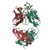 1i8mS S: Starting model for refinement |
|---|---|
| Similar structure data |


-Links
 PDBj
PDBj

- Assembly
| Deposited unit | 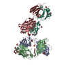
| |||||||||
|---|---|---|---|---|---|---|---|---|---|---|
| 1 | 
| |||||||||
| 2 | 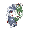
| |||||||||
| Unit cell |
| |||||||||
| Components on special symmetry positions |
|
-Components
-Antibody , 2 types, 4 molecules LAHB
| #1: Antibody | Mass: 23616.057 Da / Num. of mol.: 2 Source method: isolated from a genetically manipulated source Source: (gene. exp.) Mus musculus (house mouse) / Plasmid: PCOMB3/6-HIS / Production host:  Escherichia coli (E. coli) / Strain (production host): DH12S / References: UniProt: Q8VCP0, UniProt: I6L978*PLUS Escherichia coli (E. coli) / Strain (production host): DH12S / References: UniProt: Q8VCP0, UniProt: I6L978*PLUS#2: Antibody | Mass: 24153.158 Da / Num. of mol.: 2 Source method: isolated from a genetically manipulated source Source: (gene. exp.) Mus musculus (house mouse) / Plasmid: PCOMB3/6-HIS / Production host: Escherichia coli (E. coli) / Strain (production host): DH12S / References: UniProt: Q9JL75, UniProt: P01868*PLUS |
|---|
-Non-polymers , 6 types, 804 molecules 


| #3: Chemical | ChemComp-SO4 / Sulfate Mass: 96.063 Da / Num. of mol.: 9 / Source method: obtained synthetically / Formula: SO4 Mass: 96.063 Da / Num. of mol.: 9 / Source method: obtained synthetically / Formula: SO4#4: Chemical | HEPES Mass: 238.305 Da / Num. of mol.: 2 / Source method: obtained synthetically / Formula: C8H18N2O4S / Comment: pH buffer*YM Mass: 238.305 Da / Num. of mol.: 2 / Source method: obtained synthetically / Formula: C8H18N2O4S / Comment: pH buffer*YM#5: Chemical | ChemComp-1PE / Polyethylene glycol Mass: 238.278 Da / Num. of mol.: 4 / Source method: obtained synthetically / Formula: C10H22O6 / Comment: precipitant*YM Mass: 238.278 Da / Num. of mol.: 4 / Source method: obtained synthetically / Formula: C10H22O6 / Comment: precipitant*YM#6: Chemical | Diethylene glycol Mass: 106.120 Da / Num. of mol.: 2 / Source method: obtained synthetically / Formula: C4H10O3 Mass: 106.120 Da / Num. of mol.: 2 / Source method: obtained synthetically / Formula: C4H10O3#7: Chemical | ChemComp-GOL / Glycerol Mass: 92.094 Da / Num. of mol.: 16 / Source method: obtained synthetically / Formula: C3H8O3 Mass: 92.094 Da / Num. of mol.: 16 / Source method: obtained synthetically / Formula: C3H8O3#8: Water | ChemComp-HOH / | WaterMass: 18.015 Da / Num. of mol.: 771 / Source method: isolated from a natural source / Formula: H2O |
|---|
-Experimental details
-Experiment
| Experiment | Method: X-RAY DIFFRACTION / Number of used crystals: 1 |
|---|
- Sample preparation
| Crystal | Density Matthews: 2.69 Å3/Da / Density % sol: 54.3 % |
|---|---|
| Crystal grow | Temperature: 295 K / Method: vapor diffusion, sitting drop / pH: 8.1 Details: AMMONIUM SULFATE, HEPES, PEG 400, pH 8.1, VAPOR DIFFUSION, SITTING DROP, temperature 295K |
-Data collection
| Diffraction |
| ||||||||||||||||||
|---|---|---|---|---|---|---|---|---|---|---|---|---|---|---|---|---|---|---|---|
| Diffraction source |
| ||||||||||||||||||
| Detector |
| ||||||||||||||||||
| Radiation |
| ||||||||||||||||||
| Radiation wavelength |
| ||||||||||||||||||
| Reflection | Resolution: 1.75→48.14 Å / Num. all: 104812 / Num. obs: 98022 / % possible obs: 93.5 % / Observed criterion σ(I): 0 / Redundancy: 3.5 % / Biso Wilson estimate: 21.3 Å2 / Rmerge(I) obs: 0.044 / Net I/σ(I): 21.7 | ||||||||||||||||||
| Reflection shell | Resolution: 1.75→1.81 Å / Rmerge(I) obs: 0.487 / Mean I/σ(I) obs: 2.1 / Num. unique all: 9734 / % possible all: 94.1 |

- Processing
| Software |
| ||||||||||||||||||||||||||||||||||||
|---|---|---|---|---|---|---|---|---|---|---|---|---|---|---|---|---|---|---|---|---|---|---|---|---|---|---|---|---|---|---|---|---|---|---|---|---|---|
| Refinement | Method to determine structure: MOLECULAR REPLACEMENT Starting model: 1I8M Resolution: 1.75→48.14 Å / Rfactor Rfree error: 0.002 / Isotropic thermal model: RESTRAINED / Cross valid method: THROUGHOUT / σ(F): 0 / Stereochemistry target values: Engh & Huber
| ||||||||||||||||||||||||||||||||||||
| Solvent computation | Solvent model: FLAT MODEL / Bsol: 55.2987 Å2 / ksol: 0.373129 e/Å3 | ||||||||||||||||||||||||||||||||||||
| Displacement parameters | Biso mean: 28.5 Å2
| ||||||||||||||||||||||||||||||||||||
| Refine analyze |
| ||||||||||||||||||||||||||||||||||||
| Refinement step | Cycle: LAST / Resolution: 1.75→48.14 Å
| ||||||||||||||||||||||||||||||||||||
| Refine LS restraints |
| ||||||||||||||||||||||||||||||||||||
| LS refinement shell | Resolution: 1.75→1.81 Å / Rfactor Rfree error: 0.01 / Total num. of bins used: 10
| ||||||||||||||||||||||||||||||||||||
| Xplor file |
|
