 Movie
Movie Controller

[English] 日本語
 Yorodumi
- PDB-1p13: Crystal Structure of the Src SH2 Domain Complexed with Peptide (S... -
+ Open data

- Basic information
| Entry | Database: PDB / ID: 1p13 | ||||||
|---|---|---|---|---|---|---|---|
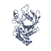
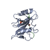
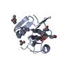
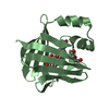
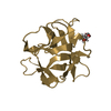
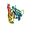
| Title | Crystal Structure of the Src SH2 Domain Complexed with Peptide (SDpYANFK) | ||||||
 Components Components |
| ||||||
 Keywords Keywords |  TRANSFERASE / TYROSINE-PROTEIN KINASE / PHOSPHORYLATION / SH3 DOMAIN TRANSFERASE / TYROSINE-PROTEIN KINASE / PHOSPHORYLATION / SH3 DOMAIN | ||||||
| Function / homology |  Function and homology information Function and homology informationSignaling by ERBB2 / Nuclear signaling by ERBB4 / PIP3 activates AKT signaling / Signaling by SCF-KIT / Regulation of KIT signaling / Signaling by EGFR / GAB1 signalosome / Regulation of gap junction activity / FCGR activation / PECAM1 interactions ...Signaling by ERBB2 / Nuclear signaling by ERBB4 / PIP3 activates AKT signaling / Signaling by SCF-KIT / Regulation of KIT signaling / Signaling by EGFR / GAB1 signalosome / Regulation of gap junction activity / FCGR activation / PECAM1 interactions / CD28 co-stimulation / CTLA4 inhibitory signaling / EPHA-mediated growth cone collapse / Ephrin signaling / G alpha (i) signalling events / GP1b-IX-V activation signalling / Recycling pathway of L1 / Thrombin signalling through proteinase activated receptors (PARs) / VEGFR2 mediated cell proliferation / RAF activation / PI5P, PP2A and IER3 Regulate PI3K/AKT Signaling / RET signaling / Receptor Mediated Mitophagy / ADP signalling through P2Y purinoceptor 1 / Downregulation of ERBB4 signaling / EPH-ephrin mediated repulsion of cells / Cyclin D associated events in G1 / Regulation of RUNX3 expression and activity / Activated NTRK3 signals through PI3K / Downstream signal transduction / MAP2K and MAPK activation / Integrin signaling / GRB2:SOS provides linkage to MAPK signaling for Integrins / MET activates PTK2 signaling / Extra-nuclear estrogen signaling / EPHB-mediated forward signaling / p130Cas linkage to MAPK signaling for integrins / VEGFA-VEGFR2 Pathway / connexin binding / osteoclast development / progesterone receptor signaling pathway / negative regulation of intrinsic apoptotic signaling pathway / bone resorption / extrinsic component of cytoplasmic side of plasma membrane / negative regulation of extrinsic apoptotic signaling pathway / non-specific protein-tyrosine kinase / non-membrane spanning protein tyrosine kinase activity / epidermal growth factor receptor signaling pathway / cell junction / protein phosphatase binding / protein tyrosine kinase activity / mitochondrial inner membrane / cell differentiation / cytoskeleton / endosome membrane / regulation of cell cycle / cell adhesion / cell cycle / phosphorylation / signaling receptor binding / focal adhesion / innate immune response / heme binding / perinuclear region of cytoplasm / protein-containing complex / ATP binding / membrane / nucleus / plasma membrane / cytosolSimilarity search - Function | ||||||
| Biological species |  Gallus gallus (chicken) Gallus gallus (chicken) | ||||||
| Method | X-RAY DIFFRACTION / SYNCHROTRON / MOLECULAR REPLACEMENT / Resolution: 1.63 Å | ||||||
 Authors Authors | Sonnenburg, E.D. / Bilwes, A. / Hunter, T. / Noel, J.P. | ||||||
 Citation Citation | Journal: Biochemistry / Year: 2003 Title: The structure of the membrane distal phosphatase domain of RPTPalpha reveals interdomain flexibility and an SH2 domain interaction region. Authors: Sonnenburg, E.D. / Bilwes, A. / Hunter, T. / Noel, J.P. | ||||||
| History |
| ||||||
| Remark 999 | SEQUENCE THERE IS NO SEQUENCE DATABASE REFERENCE FOR THE PEPTIDE CHAINS C AND D. |
- Structure visualization
| Structure viewer | Molecule: MolmilJmol/JSmol |
|---|
- Downloads & links
-Download
| PDBx/mmCIF format | 1p13.cif.gz | 59.5 KB | Display | PDBx/mmCIF format |
|---|---|---|---|---|
| PDB format | pdb1p13.ent.gz | 47.9 KB | Display | PDB format |
| PDBx/mmJSON format | 1p13.json.gz | Tree view | PDBx/mmJSON format | |
| Others |  Other downloads Other downloads |
-Validation report
| Arichive directory | https://data.pdbj.org/pub/pdb/validation_reports/p1/1p13ftp://data.pdbj.org/pub/pdb/validation_reports/p1/1p13 | HTTPS FTP |
|---|
-Related structure data

-Links
 PDBj
PDBj


- Assembly
| Deposited unit | 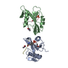
| ||||||||
|---|---|---|---|---|---|---|---|---|---|
| 1 | 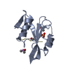
| ||||||||
| 2 | 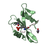
| ||||||||
| Unit cell |
|
-Components
| #1: Protein | / p60-Src / c-Src Mass: 11741.248 Da / Num. of mol.: 2 / Fragment: SH2 DOMAIN Source method: isolated from a genetically manipulated source Source: (gene. exp.) Gallus gallus (chicken) / Gene: SRC / Plasmid: pHIS8 / Species (production host): Escherichia coli / Production host:  Escherichia coli BL21(DE3) (bacteria) / Strain (production host): BL21 (DE3) / References: UniProt: P00523, EC: 2.7.1.112 Escherichia coli BL21(DE3) (bacteria) / Strain (production host): BL21 (DE3) / References: UniProt: P00523, EC: 2.7.1.112#2: Protein/peptide | Mass: 940.933 Da / Num. of mol.: 2 / Source method: obtained synthetically / Details: THE PEPTIDE WAS CHEMICALLY SYNTHESIZED. #3: Chemical | ChemComp-CAC / Cacodylic acid  Mass: 136.989 Da / Num. of mol.: 4 / Source method: obtained synthetically / Formula: C2H6AsO2 Mass: 136.989 Da / Num. of mol.: 4 / Source method: obtained synthetically / Formula: C2H6AsO2#4: Water | ChemComp-HOH / | Water Mass: 18.015 Da / Num. of mol.: 273 / Source method: isolated from a natural source / Formula: H2O Mass: 18.015 Da / Num. of mol.: 273 / Source method: isolated from a natural source / Formula: H2O |
|---|
-Experimental details
-Experiment
| Experiment | Method: X-RAY DIFFRACTION / Number of used crystals: 1 |
|---|
- Sample preparation
| Crystal | Density Matthews: 2.12 Å3/Da / Density % sol: 41.97 % | ||||||||||||||||||||||||||||||||||||
|---|---|---|---|---|---|---|---|---|---|---|---|---|---|---|---|---|---|---|---|---|---|---|---|---|---|---|---|---|---|---|---|---|---|---|---|---|---|
| Crystal grow | Temperature: 277 K / Method: vapor diffusion, hanging drop / pH: 6.5 Details: PEG 8000, sodium acetate, cacodylate, pH 6.5, VAPOR DIFFUSION, HANGING DROP, temperature 277K | ||||||||||||||||||||||||||||||||||||
| Crystal grow | *PLUS Temperature: 4 ℃ / Method: vapor diffusion, hanging drop | ||||||||||||||||||||||||||||||||||||
| Components of the solutions | *PLUS
|
-Data collection
| Diffraction | Mean temperature: 105 K |
|---|---|
| Diffraction source | Source: SYNCHROTRON / Site: SSRL  / Beamline: BL9-1 / Wavelength: 0.98 Å / Beamline: BL9-1 / Wavelength: 0.98 Å |
| Detector | Type: MACSCIENCE DIP100S / Detector: IMAGE PLATE / Date: Jun 11, 2001 |
| Radiation | Protocol: SINGLE WAVELENGTH / Monochromatic (M) / Laue (L): M / Scattering type: x-ray |
| Radiation wavelength | Wavelength: 0.98 Å / Relative weight: 1 |
| Reflection | Resolution: 1.6→42.9 Å / Num. obs: 27413 / % possible obs: 98 % / Observed criterion σ(F): 2 / Observed criterion σ(I): 2 / Biso Wilson estimate: 16.7 Å2 |
| Reflection shell | Resolution: 1.63→1.66 Å / % possible all: 69 |
| Reflection | *PLUS Num. measured all: 234548 / Rmerge(I) obs: 0.057 |
| Reflection shell | *PLUS % possible obs: 69 % / Rmerge(I) obs: 0.423 / Mean I/σ(I) obs: 2.6 |
- Processing
| Software |
| ||||||||||||||||||||||||||||||||||||
|---|---|---|---|---|---|---|---|---|---|---|---|---|---|---|---|---|---|---|---|---|---|---|---|---|---|---|---|---|---|---|---|---|---|---|---|---|---|
| Refinement | Method to determine structure: MOLECULAR REPLACEMENT / Resolution: 1.63→42.88 Å / Rfactor Rfree error: 0.005 / Isotropic thermal model: RESTRAINED / Cross valid method: THROUGHOUT / σ(F): 2 / Stereochemistry target values: Engh & Huber Details: The peptide chains, chain C and D, are discontinuous. The authors stated that the PTR residues could remain unattached from the chains.
| ||||||||||||||||||||||||||||||||||||
| Solvent computation | Solvent model: FLAT MODEL / Bsol: 45.9946 Å2 / ksol: 0.378216 e/Å3 | ||||||||||||||||||||||||||||||||||||
| Displacement parameters | Biso mean: 23.8 Å2
| ||||||||||||||||||||||||||||||||||||
| Refine analyze |
| ||||||||||||||||||||||||||||||||||||
| Refinement step | Cycle: LAST / Resolution: 1.63→42.88 Å
| ||||||||||||||||||||||||||||||||||||
| Refine LS restraints |
| ||||||||||||||||||||||||||||||||||||
| LS refinement shell | Resolution: 1.63→1.73 Å / Rfactor Rfree error: 0.018 / Total num. of bins used: 6
| ||||||||||||||||||||||||||||||||||||
| Xplor file |
| ||||||||||||||||||||||||||||||||||||
| Refinement | *PLUS Highest resolution: 1.6 Å / % reflection Rfree: 10 % / Rfactor Rfree: 0.247 / Rfactor Rwork: 0.201 | ||||||||||||||||||||||||||||||||||||
| Solvent computation | *PLUS | ||||||||||||||||||||||||||||||||||||
| Displacement parameters | *PLUS | ||||||||||||||||||||||||||||||||||||
| Refine LS restraints | *PLUS
|
