 Movie
Movie Controller


+ Open data

- Basic information
| Entry | Database: PDB / ID: 1p0q | |||||||||
|---|---|---|---|---|---|---|---|---|---|---|
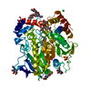
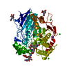
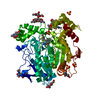
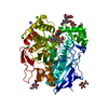
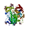
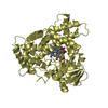
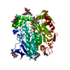
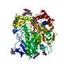
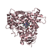
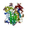
| Title | Crystal structure of soman-aged human butyryl cholinesterase | |||||||||
 Components Components | Cholinesterase | |||||||||
 Keywords Keywords | HYDROLASE / Serine hydrolase / organophosphates / soman / cholinesterase / conformational change | |||||||||
| Function / homology |  Function and homology informationcholinesterase / cocaine metabolic process / neuroblast differentiation / Neurotransmitter clearance / choline metabolic process / choline binding / response to folic acid / response to alkaloid / acetylcholine catabolic process / negative regulation of synaptic transmission ...cholinesterase / cocaine metabolic process / neuroblast differentiation / Neurotransmitter clearance / choline metabolic process / choline binding / response to folic acid / response to alkaloid / acetylcholine catabolic process / negative regulation of synaptic transmission / cholinesterase activity / peptide hormone processing / acetylcholinesterase activity / hydrolase activity, acting on ester bonds / nuclear envelope lumen / Aspirin ADME / Synthesis of PC / Synthesis, secretion, and deacylation of Ghrelin / catalytic activity / response to glucocorticoid / xenobiotic metabolic process / learning / amyloid-beta binding / blood microparticle / negative regulation of cell population proliferation / endoplasmic reticulum lumen / enzyme binding / extracellular space / extracellular region / identical protein binding / plasma membrane Function and homology informationcholinesterase / cocaine metabolic process / neuroblast differentiation / Neurotransmitter clearance / choline metabolic process / choline binding / response to folic acid / response to alkaloid / acetylcholine catabolic process / negative regulation of synaptic transmission ...cholinesterase / cocaine metabolic process / neuroblast differentiation / Neurotransmitter clearance / choline metabolic process / choline binding / response to folic acid / response to alkaloid / acetylcholine catabolic process / negative regulation of synaptic transmission / cholinesterase activity / peptide hormone processing / acetylcholinesterase activity / hydrolase activity, acting on ester bonds / nuclear envelope lumen / Aspirin ADME / Synthesis of PC / Synthesis, secretion, and deacylation of Ghrelin / catalytic activity / response to glucocorticoid / xenobiotic metabolic process / learning / amyloid-beta binding / blood microparticle / negative regulation of cell population proliferation / endoplasmic reticulum lumen / enzyme binding / extracellular space / extracellular region / identical protein binding / plasma membraneSimilarity search - Function | |||||||||
| Biological species |  Homo sapiens (human) Homo sapiens (human) | |||||||||
| Method | X-RAY DIFFRACTION / SYNCHROTRON / MOLECULAR REPLACEMENT / Resolution: 2.43 Å | |||||||||
 Authors Authors | Nicolet, Y. / Lockridge, O. / Masson, P. / Fontecilla-Camps, J.C. / Nachon, F. | |||||||||
 Citation Citation | Journal: J.Biol.Chem. / Year: 2003 Title: Crystal structure of human butyrylcholinesterase and of its complexes with substrate and products. Authors: Nicolet, Y. / Lockridge, O. / Masson, P. / Fontecilla-Camps, J.C. / Nachon, F. #1: Journal: Eur.J.Biochem. / Year: 2002Title: Engineering of a monomeric and low-glycosylated form of human butyrylcholinesterase: expression, purification, characterization and crystallization Authors: Nachon, F. / Nicolet, Y. / Viguie, N. / Masson, P. / Fontecilla-Camps, J.C. / Lockridge, O. | |||||||||
| History |
|
- Structure visualization
| Structure viewer | Molecule: MolmilJmol/JSmol |
|---|
- Downloads & links
-Download
| PDBx/mmCIF format | 1p0q.cif.gz | 129.2 KB | Display | PDBx/mmCIF format |
|---|---|---|---|---|
| PDB format | pdb1p0q.ent.gz | 99.1 KB | Display | PDB format |
| PDBx/mmJSON format | 1p0q.json.gz | Tree view | PDBx/mmJSON format | |
| Others |  Other downloads Other downloads |
-Validation report
| Arichive directory | https://data.pdbj.org/pub/pdb/validation_reports/p0/1p0qftp://data.pdbj.org/pub/pdb/validation_reports/p0/1p0q | HTTPS FTP |
|---|
-Related structure data
| Related structure data |  1p0iSC 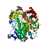 1p0mC  1p0pC S: Starting model for refinement C: citing same article ( |
|---|---|
| Similar structure data |
-Links
 PDBj
PDBj


- Assembly
| Deposited unit | 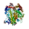
| ||||||||
|---|---|---|---|---|---|---|---|---|---|
| 1 |
| ||||||||
| Unit cell |
| ||||||||
| Components on special symmetry positions |
|
-Components
-Protein , 1 types, 1 molecules A
| #1: Protein | / Acylcholine acylhydrolase / Choline esterase II / Butyrylcholine esterase / Pseudocholinesterase Mass: 59713.512 Da / Num. of mol.: 1 / Mutation: N17Q, N455Q, N481Q, N486Q Source method: isolated from a genetically manipulated source Source: (gene. exp.) Homo sapiens (human) / Gene: BCHE OR CHE1 / Cell line (production host): ovary cells / Production host:   Cricetulus griseus (Chinese hamster) / References: UniProt: P06276, cholinesterase Cricetulus griseus (Chinese hamster) / References: UniProt: P06276, cholinesterase |
|---|
-Sugars , 2 types, 4 molecules 
| #2: Polysaccharide | / Mass: 570.542 Da / Num. of mol.: 2 / Source method: obtained synthetically| #3: Sugar | N-Acetylglucosamine Type: D-saccharide, beta linking / Mass: 221.208 Da / Num. of mol.: 2 / Source method: obtained synthetically / Formula: C8H15NO6 Type: D-saccharide, beta linking / Mass: 221.208 Da / Num. of mol.: 2 / Source method: obtained synthetically / Formula: C8H15NO6 |
|---|
-Non-polymers , 5 types, 291 molecules 


| #4: Chemical | ChemComp-SO4 / Sulfate Mass: 96.063 Da / Num. of mol.: 1 / Source method: obtained synthetically / Formula: SO4 Mass: 96.063 Da / Num. of mol.: 1 / Source method: obtained synthetically / Formula: SO4 | ||
|---|---|---|---|
| #5: Chemical | ChemComp-CL / Chloride Mass: 35.453 Da / Num. of mol.: 1 / Source method: obtained synthetically / Formula: Cl Mass: 35.453 Da / Num. of mol.: 1 / Source method: obtained synthetically / Formula: Cl | ||
| #6: Chemical | ChemComp-VXA / Methylphosphinic acid Mass: 79.015 Da / Num. of mol.: 1 / Source method: obtained synthetically / Formula: CH4O2P Mass: 79.015 Da / Num. of mol.: 1 / Source method: obtained synthetically / Formula: CH4O2P | ||
| #7: Chemical | Glycerol Mass: 92.094 Da / Num. of mol.: 2 / Source method: obtained synthetically / Formula: C3H8O3 Mass: 92.094 Da / Num. of mol.: 2 / Source method: obtained synthetically / Formula: C3H8O3#8: Water | ChemComp-HOH / | WaterMass: 18.015 Da / Num. of mol.: 286 / Source method: isolated from a natural source / Formula: H2O |
-Experimental details
-Experiment
| Experiment | Method: X-RAY DIFFRACTION / Number of used crystals: 1 |
|---|
- Sample preparation
| Crystal | Density Matthews: 2.97 Å3/Da / Density % sol: 58.31 % | ||||||||||||||||||||||||
|---|---|---|---|---|---|---|---|---|---|---|---|---|---|---|---|---|---|---|---|---|---|---|---|---|---|
| Crystal grow | Temperature: 298 K / Method: vapor diffusion, hanging drop / pH: 8 Details: ammonium sulfate, Tris-(hydroxymethyl)-aminomethane, pH 8.0, VAPOR DIFFUSION, HANGING DROP, temperature 298.0K | ||||||||||||||||||||||||
| Crystal grow | *PLUS Temperature: 20 ℃ / pH: 6.5 / Method: vapor diffusion, hanging drop / Details: Nachon, F., (2002) Eur.J.Biochem., 269, 630. | ||||||||||||||||||||||||
| Components of the solutions | *PLUS
|
-Data collection
| Diffraction | Mean temperature: 100 K |
|---|---|
| Diffraction source | Source: SYNCHROTRON / Site: ESRF  / Beamline: ID14-1 / Wavelength: 0.934 Å / Beamline: ID14-1 / Wavelength: 0.934 Å |
| Detector | Type: ADSC QUANTUM 4 / Detector: CCD |
| Radiation | Protocol: SINGLE WAVELENGTH / Monochromatic (M) / Laue (L): M / Scattering type: x-ray |
| Radiation wavelength | Wavelength: 0.934 Å / Relative weight: 1 |
| Reflection | Resolution: 2.43→41.52 Å / Num. all: 26509 / Num. obs: 25868 / % possible obs: 90.8 % / Observed criterion σ(F): 0 / Observed criterion σ(I): 1 / Redundancy: 10.2 % / Biso Wilson estimate: 39.1 Å2 / Rsym value: 0.071 / Net I/σ(I): 7.6 |
| Reflection shell | Resolution: 2.43→2.56 Å / Redundancy: 6.2 % / Mean I/σ(I) obs: 3.3 / Num. unique all: 3723 / Rsym value: 0.217 / % possible all: 88.6 |
| Reflection | *PLUS Lowest resolution: 40.6 Å / Rmerge(I) obs: 0.071 |
| Reflection shell | *PLUS % possible obs: 88.6 % / Rmerge(I) obs: 0.217 |
- Processing
| Software |
| ||||||||||||||||||||||||||||||||||||
|---|---|---|---|---|---|---|---|---|---|---|---|---|---|---|---|---|---|---|---|---|---|---|---|---|---|---|---|---|---|---|---|---|---|---|---|---|---|
| Refinement | Method to determine structure: MOLECULAR REPLACEMENT Starting model: PDB ENTRY 1P0I Resolution: 2.43→40.64 Å / Rfactor Rfree error: 0.007 / Isotropic thermal model: RESTRAINED / Cross valid method: THROUGHOUT / σ(F): 0 / Stereochemistry target values: Engh & Huber
| ||||||||||||||||||||||||||||||||||||
| Solvent computation | Solvent model: FLAT MODEL / Bsol: 85.9698 Å2 / ksol: 0.37042 e/Å3 | ||||||||||||||||||||||||||||||||||||
| Displacement parameters | Biso mean: 38.3 Å2
| ||||||||||||||||||||||||||||||||||||
| Refine analyze |
| ||||||||||||||||||||||||||||||||||||
| Refinement step | Cycle: LAST / Resolution: 2.43→40.64 Å
| ||||||||||||||||||||||||||||||||||||
| Refine LS restraints |
| ||||||||||||||||||||||||||||||||||||
| LS refinement shell | Resolution: 2.43→2.58 Å / Rfactor Rfree error: 0.021 / Total num. of bins used: 6
| ||||||||||||||||||||||||||||||||||||
| Xplor file |
| ||||||||||||||||||||||||||||||||||||
| Refinement | *PLUS Lowest resolution: 40.6 Å | ||||||||||||||||||||||||||||||||||||
| Solvent computation | *PLUS | ||||||||||||||||||||||||||||||||||||
| Displacement parameters | *PLUS | ||||||||||||||||||||||||||||||||||||
| Refine LS restraints | *PLUS
|
