 Movie
Movie Controller

[English] 日本語
 Yorodumi
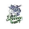
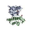
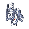
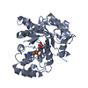
- PDB-1oyf: Crystal Structure of Russelles viper (Daboia russellii pulchella)... -
+ Open data

- Basic information
| Entry | Database: PDB / ID: 1oyf | ||||||
|---|---|---|---|---|---|---|---|
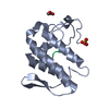
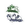
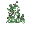
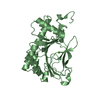
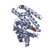
| Title | Crystal Structure of Russelles viper (Daboia russellii pulchella) phospholipase A2 in a complex with venom 6-methyl heptanol | ||||||
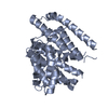
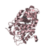
 Components Components | (Phospholipase ... ) x 2 ) x 2 | ||||||
 Keywords Keywords | HYDROLASE / Phospholipase A2 / complex / Catalysis / Inhibition | ||||||
| Function / homology |  Function and homology information Function and homology informationcalcium-dependent phospholipase A2 activity / phospholipase A2 / arachidonic acid secretion / phospholipid metabolic process / lipid catabolic process / negative regulation of T cell proliferation / phospholipid binding / toxin activity / calcium ion binding / extracellular regionSimilarity search - Function | ||||||
| Biological species |  Daboia russellii pulchella (snake) Daboia russellii pulchella (snake) | ||||||
| Method | X-RAY DIFFRACTION / MOLECULAR REPLACEMENT / Resolution: 2.45 Å | ||||||
 Authors Authors | Singh, N. / Jabeen, T. / Sharma, S. / Singh, T.P. | ||||||
 Citation Citation | Journal: To be Published Title: Crystal Structure of Russelles viper (Daboia russellii pulchella) phospholipase A2 in a complex with venom 6-methyl heptanol Authors: Singh, N. / Jabeen, T. / Sharma, S. / Singh, T.P. | ||||||
| History |
|
- Structure visualization
| Structure viewer | Molecule: MolmilJmol/JSmol |
|---|
- Downloads & links
-Download
| PDBx/mmCIF format | 1oyf.cif.gz | 65.5 KB | Display | PDBx/mmCIF format |
|---|---|---|---|---|
| PDB format | pdb1oyf.ent.gz | 47.6 KB | Display | PDB format |
| PDBx/mmJSON format | 1oyf.json.gz | Tree view | PDBx/mmJSON format | |
| Others |  Other downloads Other downloads |
-Validation report
| Arichive directory | https://data.pdbj.org/pub/pdb/validation_reports/oy/1oyfftp://data.pdbj.org/pub/pdb/validation_reports/oy/1oyf | HTTPS FTP |
|---|
-Related structure data
| Related structure data | 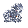 1fvoS S: Starting model for refinement |
|---|---|
| Similar structure data |
-Links
 PDBj
PDBj


- Assembly
| Deposited unit | 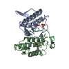
| ||||||||
|---|---|---|---|---|---|---|---|---|---|
| 1 |
| ||||||||
| Unit cell |
|
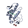
-Components
-Phospholipase ... , 2 types, 2 molecules AB
| #1: Protein | Mass: 13656.729 Da / Num. of mol.: 1 / Source method: isolated from a natural source / Source: (natural) Daboia russellii pulchella (snake) / Secretion: Venom / Species: russellii / Strain: pulchella / References: UniProt: P59071, phospholipase A2 |
|---|---|
| #2: Protein | Mass: 13657.780 Da / Num. of mol.: 1 / Source method: isolated from a natural source / Source: (natural) Daboia russellii pulchella (snake) / Secretion: Venom / Species: russellii / Strain: pulchella / References: phospholipase A2 |
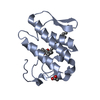
-Non-polymers , 4 types, 152 molecules 


| #3: Chemical | ChemComp-SO4 / Sulfate Mass: 96.063 Da / Num. of mol.: 1 / Source method: obtained synthetically / Formula: SO4 Mass: 96.063 Da / Num. of mol.: 1 / Source method: obtained synthetically / Formula: SO4 | ||||
|---|---|---|---|---|---|
| #4: Chemical |  Mass: 130.228 Da / Num. of mol.: 2 / Source method: obtained synthetically / Formula: C8H18O Mass: 130.228 Da / Num. of mol.: 2 / Source method: obtained synthetically / Formula: C8H18O#5: Chemical | ChemComp-ACY / | Acetic acid Mass: 60.052 Da / Num. of mol.: 1 / Source method: obtained synthetically / Formula: C2H4O2 Mass: 60.052 Da / Num. of mol.: 1 / Source method: obtained synthetically / Formula: C2H4O2#6: Water | ChemComp-HOH / | WaterMass: 18.015 Da / Num. of mol.: 148 / Source method: isolated from a natural source / Formula: H2O |
-Experimental details
-Experiment
| Experiment | Method: X-RAY DIFFRACTION / Number of used crystals: 1 |
|---|
- Sample preparation
| Crystal | Density Matthews: 2.19 Å3/Da / Density % sol: 43.9 % |
|---|---|
| Crystal grow | Temperature: 298 K / Method: vapor diffusion, hanging drop / pH: 6.5 Details: 0.1M Cacodylate buffer, 0.2M Ammonoum Sulphate, 15% PEG8000, pH 6.5, VAPOR DIFFUSION, HANGING DROP, temperature 298K |
-Data collection
| Diffraction | Mean temperature: 291 K |
|---|---|
| Diffraction source | Source: ROTATING ANODE / Type: RIGAKU RU200 / Wavelength: 1.5418 Å |
| Detector | Type: MARRESEARCH / Detector: IMAGE PLATE / Date: Sep 4, 2002 / Details: Monochromator |
| Radiation | Monochromator: Graphite / Protocol: SINGLE WAVELENGTH / Monochromatic (M) / Laue (L): M / Scattering type: x-ray |
| Radiation wavelength | Wavelength: 1.5418 Å / Relative weight: 1 |
| Reflection | Resolution: 2.4→20 Å / Num. all: 58251 / Num. obs: 7984 / % possible obs: 98.1 % / Observed criterion σ(F): 0 / Observed criterion σ(I): 0 / Redundancy: 7.3 % / Biso Wilson estimate: 18.9 Å2 / Rsym value: 0.072 / Net I/σ(I): 25.2 |
| Reflection shell | Resolution: 2.4→2.5 Å / Mean I/σ(I) obs: 3.2 / Rsym value: 0.31 / % possible all: 95.3 |
- Processing
| Software |
| ||||||||||||||||||||||||||||||||||||
|---|---|---|---|---|---|---|---|---|---|---|---|---|---|---|---|---|---|---|---|---|---|---|---|---|---|---|---|---|---|---|---|---|---|---|---|---|---|
| Refinement | Method to determine structure: MOLECULAR REPLACEMENT Starting model: 1FVO Resolution: 2.45→19.86 Å / Rfactor Rfree error: 0.012 / Data cutoff high absF: 173506.47 / Data cutoff low absF: 0 / Isotropic thermal model: RESTRAINED / Cross valid method: THROUGHOUT / σ(F): 0 / σ(I): 0 / Stereochemistry target values: Engh & Huber Details: Trp31 im molecule A is having double positions. Both were refined separately. The distance between the C of GLY A 30 and alternate conformation B of TRP A 31 is long.
| ||||||||||||||||||||||||||||||||||||
| Solvent computation | Solvent model: FLAT MODEL / Bsol: 40.0431 Å2 / ksol: 0.347483 e/Å3 | ||||||||||||||||||||||||||||||||||||
| Displacement parameters | Biso mean: 27.3 Å2 | ||||||||||||||||||||||||||||||||||||
| Refine analyze |
| ||||||||||||||||||||||||||||||||||||
| Refinement step | Cycle: LAST / Resolution: 2.45→19.86 Å
| ||||||||||||||||||||||||||||||||||||
| Refine LS restraints |
| ||||||||||||||||||||||||||||||||||||
| LS refinement shell | Resolution: 2.44→2.59 Å / Rfactor Rfree error: 0.05 / Total num. of bins used: 6
| ||||||||||||||||||||||||||||||||||||
| Xplor file |
|
