 Movie
Movie Controller


+ Open data

- Basic information
| Entry | Database: PDB / ID: 1otv | ||||||
|---|---|---|---|---|---|---|---|
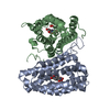
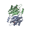
| Title | PqqC, Pyrroloquinolinquinone Synthase C | ||||||
 Components Components | Coenzyme PQQ synthesis protein C | ||||||
 Keywords Keywords |  BIOSYNTHETIC PROTEIN / seven helix bundle / dimer / PQQ biosynthesis enzyme BIOSYNTHETIC PROTEIN / seven helix bundle / dimer / PQQ biosynthesis enzyme | ||||||
| Function / homology |  Function and homology informationpyrroloquinoline-quinone synthase / pyrroloquinoline-quinone synthase activity / pyrroloquinoline quinone biosynthetic process / : / sulfur compound metabolic process Function and homology informationpyrroloquinoline-quinone synthase / pyrroloquinoline-quinone synthase activity / pyrroloquinoline quinone biosynthetic process / : / sulfur compound metabolic processSimilarity search - Function | ||||||
| Biological species |  Klebsiella pneumoniae (bacteria) Klebsiella pneumoniae (bacteria) | ||||||
| Method | X-RAY DIFFRACTION / SYNCHROTRON / MOLECULAR REPLACEMENT / Resolution: 2.1 Å | ||||||
 Authors Authors | Magnusson, O.T. / Toyama, H. / Saeki, M. / Rojas, A. / Reed, J.C. / Adachi, O. / Klinman, J.P. / SChwarzenbacher, R. | ||||||
 Citation Citation | Journal: Proc.Natl.Acad.Sci.USA / Year: 2004 Title: Quinone Biogenesis: Structure and Mechanism of PqqC, the Final Catalyst in the Production of Pyrroloquinoline Quinone. Authors: Magnusson, O.T. / Toyama, H. / Saeki, M. / Rojas, A. / Reed, J.C. / Adachi, O. / Liddington, R.C. / Klinman, J.P. / Schwarzenbacher, R. | ||||||
| History |
| ||||||
| Remark 650 | author determined the secondary structure |
- Structure visualization
| Structure viewer | Molecule: MolmilJmol/JSmol |
|---|
- Downloads & links
-Download
| PDBx/mmCIF format | 1otv.cif.gz | 115.5 KB | Display | PDBx/mmCIF format |
|---|---|---|---|---|
| PDB format | pdb1otv.ent.gz | 91.3 KB | Display | PDB format |
| PDBx/mmJSON format | 1otv.json.gz | Tree view | PDBx/mmJSON format | |
| Others |  Other downloads Other downloads |
-Validation report
| Arichive directory | https://data.pdbj.org/pub/pdb/validation_reports/ot/1otvftp://data.pdbj.org/pub/pdb/validation_reports/ot/1otv | HTTPS FTP |
|---|
-Related structure data


-Links
 PDBj
PDBj- Assembly
| Deposited unit | 
| ||||||||
|---|---|---|---|---|---|---|---|---|---|
| 1 |
| ||||||||
| 2 | 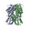
| ||||||||
| Unit cell |
| ||||||||
| Details | The biological assembly is a dimer |
-Components
| #1: Protein | Mass: 30142.113 Da / Num. of mol.: 2 / Mutation: A21D Source method: isolated from a genetically manipulated source Source: (gene. exp.) Klebsiella pneumoniae (bacteria) / Gene: PQQC / Plasmid: pET101 / Species (production host): Escherichia coli / Production host: Escherichia coli BL21 (bacteria) / Strain (production host): BL21 / References: UniProt: P27505#2: Water | ChemComp-HOH / | Water Mass: 18.015 Da / Num. of mol.: 130 / Source method: isolated from a natural source / Formula: H2O Mass: 18.015 Da / Num. of mol.: 130 / Source method: isolated from a natural source / Formula: H2O |
|---|
-Experimental details
-Experiment
| Experiment | Method: X-RAY DIFFRACTION / Number of used crystals: 1 |
|---|
- Sample preparation
| Crystal | Density Matthews: 2.51 Å3/Da / Density % sol: 50.68 % |
|---|---|
| Crystal grow | Temperature: 298 K / Method: vapor diffusion, sitting drop / pH: 6.8 Details: 1.4M ammonium sulphate, cacodylate , pH 6.8, VAPOR DIFFUSION, SITTING DROP, temperature 25K |
-Data collection
| Diffraction | Mean temperature: 100 K |
|---|---|
| Diffraction source | Source: SYNCHROTRON / Site: SSRL  / Beamline: BL9-1 / Wavelength: 0.978 Å / Beamline: BL9-1 / Wavelength: 0.978 Å |
| Detector | Type: ADSC QUANTUM 4 / Detector: CCD / Date: May 1, 2002 |
| Radiation | Protocol: MAD / Monochromatic (M) / Laue (L): M / Scattering type: x-ray |
| Radiation wavelength | Wavelength: 0.978 Å / Relative weight: 1 |
| Reflection | Resolution: 2.1→69 Å / Num. all: 35745 / Num. obs: 35743 / % possible obs: 98.1 % / Observed criterion σ(F): 0 / Observed criterion σ(I): 0 / Redundancy: 4.4 % / Biso Wilson estimate: 38.2 Å2 / Rsym value: 0.145 / Net I/σ(I): 10.1 |
| Reflection shell | Resolution: 2.1→2.155 Å / Redundancy: 3.6 % / Mean I/σ(I) obs: 2.3 / Num. unique all: 1787 / Rsym value: 0.41 / % possible all: 96.2 |
- Processing
| Software |
| ||||||||||||||||||||||||||||||||||||||||||||||||||||||||||||||||||||||||||||||||||||||||||||||||||||
|---|---|---|---|---|---|---|---|---|---|---|---|---|---|---|---|---|---|---|---|---|---|---|---|---|---|---|---|---|---|---|---|---|---|---|---|---|---|---|---|---|---|---|---|---|---|---|---|---|---|---|---|---|---|---|---|---|---|---|---|---|---|---|---|---|---|---|---|---|---|---|---|---|---|---|---|---|---|---|---|---|---|---|---|---|---|---|---|---|---|---|---|---|---|---|---|---|---|---|---|---|---|
| Refinement | Method to determine structure: MOLECULAR REPLACEMENT / Resolution: 2.1→69.01 Å / Cor.coef. Fo:Fc: 0.953 / Cor.coef. Fo:Fc free: 0.936 / SU B: 4.518 / SU ML: 0.118 / TLS residual ADP flag: LIKELY RESIDUAL / Isotropic thermal model: Isotropic / Cross valid method: THROUGHOUT / σ(F): 0 / σ(I): 0 / ESU R: 0.2 / ESU R Free: 0.17 / Stereochemistry target values: MAXIMUM LIKELIHOOD Details: Residues 152-160 of chain A and 151-162 of chain B are disordered, with an occupancy of 0. HYDROGENS HAVE BEEN ADDED IN THE RIDING POSITIONS
| ||||||||||||||||||||||||||||||||||||||||||||||||||||||||||||||||||||||||||||||||||||||||||||||||||||
| Solvent computation | Ion probe radii: 0.8 Å / Shrinkage radii: 0.8 Å / VDW probe radii: 1.4 Å / Solvent model: BABINET MODEL WITH MASK | ||||||||||||||||||||||||||||||||||||||||||||||||||||||||||||||||||||||||||||||||||||||||||||||||||||
| Displacement parameters | Biso mean: 32.916 Å2
| ||||||||||||||||||||||||||||||||||||||||||||||||||||||||||||||||||||||||||||||||||||||||||||||||||||
| Refine analyze | Luzzati coordinate error obs: 0.136 Å | ||||||||||||||||||||||||||||||||||||||||||||||||||||||||||||||||||||||||||||||||||||||||||||||||||||
| Refinement step | Cycle: LAST / Resolution: 2.1→69.01 Å
| ||||||||||||||||||||||||||||||||||||||||||||||||||||||||||||||||||||||||||||||||||||||||||||||||||||
| Refine LS restraints |
| ||||||||||||||||||||||||||||||||||||||||||||||||||||||||||||||||||||||||||||||||||||||||||||||||||||
| LS refinement shell | Resolution: 2.1→2.155 Å / Total num. of bins used: 20
| ||||||||||||||||||||||||||||||||||||||||||||||||||||||||||||||||||||||||||||||||||||||||||||||||||||
| Refinement TLS params. | Method: refined / Origin x: 33.957 Å / Origin y: 41.794 Å / Origin z: 17.269 Å
| ||||||||||||||||||||||||||||||||||||||||||||||||||||||||||||||||||||||||||||||||||||||||||||||||||||
| Refinement TLS group |
|
