 Movie
Movie Controller


+ Open data

- Basic information
| Entry | Database: PDB / ID: 1oqw | ||||||
|---|---|---|---|---|---|---|---|
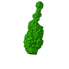
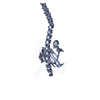
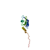
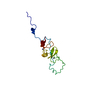
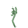
| Title | Full-Length PAK Pilin from Pseudomonas aeruginosa | ||||||
 Components Components | Fimbrial protein | ||||||
 Keywords Keywords |  CELL ADHESION / Type IV pilin / fiber-forming protein / adhesion / Pseudomonas aerugionosa / PAK pilin CELL ADHESION / Type IV pilin / fiber-forming protein / adhesion / Pseudomonas aerugionosa / PAK pilin | ||||||
| Function / homology |  Function and homology information Function and homology information | ||||||
| Biological species |   Pseudomonas aeruginosa (bacteria) Pseudomonas aeruginosa (bacteria) | ||||||
| Method | X-RAY DIFFRACTION / SYNCHROTRON / MOLECULAR REPLACEMENT / Resolution: 2 Å | ||||||
 Authors Authors | Craig, L. / Arvai, A.S. / Forest, K.T. / Tainer, J.A. | ||||||
 Citation Citation | Journal: Mol.Cell / Year: 2003 Title: Type IV Pilin Structure and Assembly: X-Ray and EM Analyses of Vibrio cholerae Toxin-Coregulated Pilus and Pseudomonas aeruginosa PAK Pilin Authors: Craig, L. / Taylor, R.K. / Pique, M.E. / Adair, B.A. / Arvai, A.S. / Singh, M. / Lloyd, S.J. / Shin, D.S. / Getzoff, E.D. / Yeager, M. / Forest, K.T. / Tainer, J.A. | ||||||
| History |
|
- Structure visualization
| Structure viewer | Molecule: MolmilJmol/JSmol |
|---|
- Downloads & links
-Download
| PDBx/mmCIF format | 1oqw.cif.gz | 69.7 KB | Display | PDBx/mmCIF format |
|---|---|---|---|---|
| PDB format | pdb1oqw.ent.gz | 51.9 KB | Display | PDB format |
| PDBx/mmJSON format | 1oqw.json.gz | Tree view | PDBx/mmJSON format | |
| Others |  Other downloads Other downloads |
-Validation report
| Arichive directory | https://data.pdbj.org/pub/pdb/validation_reports/oq/1oqwftp://data.pdbj.org/pub/pdb/validation_reports/oq/1oqw | HTTPS FTP |
|---|
-Related structure data
| Related structure data |  1oqvC  1dzoS C: citing same article ( S: Starting model for refinement |
|---|---|
| Similar structure data |


-Links
 PDBj
PDBj
- Assembly
| Deposited unit | 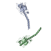
| ||||||||
|---|---|---|---|---|---|---|---|---|---|
| 1 | 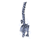
| ||||||||
| 2 | 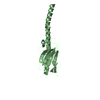
| ||||||||
| Unit cell |
| ||||||||
| Details | thousands of pilin subunits assemble to form a long thin filament ~8 nm in diameter and several microns in length |
-Components
| #1: Protein | Mass: 15021.180 Da / Num. of mol.: 2 / Source method: isolated from a natural source / Source: (natural) Pseudomonas aeruginosa (bacteria) / Strain: K / References: UniProt: P02973#2: Water | ChemComp-HOH / | Water Mass: 18.015 Da / Num. of mol.: 282 / Source method: isolated from a natural source / Formula: H2O Mass: 18.015 Da / Num. of mol.: 282 / Source method: isolated from a natural source / Formula: H2O |
|---|
-Experimental details
-Experiment
| Experiment | Method: X-RAY DIFFRACTION / Number of used crystals: 1 |
|---|
- Sample preparation
| Crystal | Density Matthews: 3.21 Å3/Da / Density % sol: 61.38 % | ||||||||||||||||||||||||||||||||||||||||||
|---|---|---|---|---|---|---|---|---|---|---|---|---|---|---|---|---|---|---|---|---|---|---|---|---|---|---|---|---|---|---|---|---|---|---|---|---|---|---|---|---|---|---|---|
| Crystal grow | Temperature: 293 K / Method: vapor diffusion, hanging drop / pH: 5.6 Details: 15% MPD, 35% PEG 4000, 100 mM sodium citrate, 2.5 mM MnCl2, pH 5.6, VAPOR DIFFUSION, HANGING DROP, temperature 293K | ||||||||||||||||||||||||||||||||||||||||||
| Crystal grow | *PLUS Method: vapor diffusion, sitting drop | ||||||||||||||||||||||||||||||||||||||||||
| Components of the solutions | *PLUS
|
-Data collection
| Diffraction | Mean temperature: 100 K |
|---|---|
| Diffraction source | Source: SYNCHROTRON / Site: SSRL  / Beamline: BL9-2 / Wavelength: 0.9791 Å / Beamline: BL9-2 / Wavelength: 0.9791 Å |
| Detector | Type: ADSC QUANTUM 4 / Detector: CCD / Date: Jun 28, 2000 |
| Radiation | Monochromator: double crystal monochromator / Protocol: SINGLE WAVELENGTH / Monochromatic (M) / Laue (L): M / Scattering type: x-ray |
| Radiation wavelength | Wavelength: 0.9791 Å / Relative weight: 1 |
| Reflection | Resolution: 2→30 Å / Num. all: 29079 / Num. obs: 28468 / % possible obs: 97.2 % / Observed criterion σ(F): 0 / Observed criterion σ(I): -3.7 / Redundancy: 3.24 % / Biso Wilson estimate: 33.7 Å2 / Rmerge(I) obs: 0.079 / Net I/σ(I): 14.8 |
| Reflection shell | Resolution: 2→2.07 Å / Redundancy: 2.28 % / Rmerge(I) obs: 0.33 / Mean I/σ(I) obs: 2.4 / Num. unique all: 2875 / % possible all: 83.6 |
| Reflection | *PLUS Num. obs: 29308 / Num. measured all: 324152 |
| Reflection shell | *PLUS % possible obs: 83.6 % / Rmerge(I) obs: 0.329 |
- Processing
| Software |
| |||||||||||||||||||||||||
|---|---|---|---|---|---|---|---|---|---|---|---|---|---|---|---|---|---|---|---|---|---|---|---|---|---|---|
| Refinement | Method to determine structure: MOLECULAR REPLACEMENT Starting model: 1DZO.pdb Resolution: 2→30 Å / Cross valid method: used throughout refinement / σ(F): 0 / Stereochemistry target values: Engh & Huber
| |||||||||||||||||||||||||
| Displacement parameters |
| |||||||||||||||||||||||||
| Refine analyze | Luzzati coordinate error obs: 0.25 Å / Luzzati d res low obs: 5 Å | |||||||||||||||||||||||||
| Refinement step | Cycle: LAST / Resolution: 2→30 Å
| |||||||||||||||||||||||||
| Refine LS restraints |
| |||||||||||||||||||||||||
| LS refinement shell | Resolution: 2→2.3 Å /
| |||||||||||||||||||||||||
| Refinement | *PLUS Highest resolution: 2 Å / Lowest resolution: 30 Å / % reflection Rfree: 5 % | |||||||||||||||||||||||||
| Solvent computation | *PLUS | |||||||||||||||||||||||||
| Displacement parameters | *PLUS | |||||||||||||||||||||||||
| Refine LS restraints | *PLUS Type: c_bond_d / Dev ideal: 0.007 |
