 Movie
Movie Controller

[English] 日本語
 Yorodumi
- PDB-1oqr: Crystal structure of C73S mutant of putidaredoxin, a [2Fe-2S] fer... -
+ Open data

- Basic information
| Entry | Database: PDB / ID: 1oqr | ||||||
|---|---|---|---|---|---|---|---|
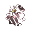
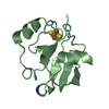
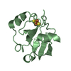
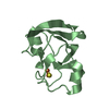
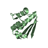
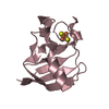
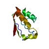
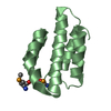
| Title | Crystal structure of C73S mutant of putidaredoxin, a [2Fe-2S] ferredoxin from Pseudomonas putida, at 1.65A resolution | ||||||
 Components Components | Putidaredoxin | ||||||
 Keywords Keywords |  ELECTRON TRANSPORT / electron transfer ELECTRON TRANSPORT / electron transfer | ||||||
| Function / homology |  Function and homology information Function and homology informationP450-containing electron transport chain / 2 iron, 2 sulfur cluster binding / metal ion binding / cytosolSimilarity search - Function | ||||||
| Biological species |  Pseudomonas putida (bacteria) Pseudomonas putida (bacteria) | ||||||
| Method | X-RAY DIFFRACTION / SYNCHROTRON / MOLECULAR REPLACEMENT / Resolution: 1.65 Å | ||||||
 Authors Authors | Sevrioukova, I.F. / Garcia, C. / Li, H. / Bhaskar, B. / Poulos, T.L. | ||||||
 Citation Citation | Journal: J.MOL.BIOL. / Year: 2003 Title: Crystal structure of putidaredoxin, the [2Fe-2S] component of the P450cam monooxygenase system from Pseudomonas putida Authors: Sevrioukova, I.F. / Garcia, C. / Li, H. / Bhaskar, B. / Poulos, T.L. | ||||||
| History |
|
- Structure visualization
| Structure viewer | Molecule: MolmilJmol/JSmol |
|---|
- Downloads & links
-Download
| PDBx/mmCIF format | 1oqr.cif.gz | 74.5 KB | Display | PDBx/mmCIF format |
|---|---|---|---|---|
| PDB format | pdb1oqr.ent.gz | 55.8 KB | Display | PDB format |
| PDBx/mmJSON format | 1oqr.json.gz | Tree view | PDBx/mmJSON format | |
| Others |  Other downloads Other downloads |
-Validation report
| Arichive directory | https://data.pdbj.org/pub/pdb/validation_reports/oq/1oqrftp://data.pdbj.org/pub/pdb/validation_reports/oq/1oqr | HTTPS FTP |
|---|
-Related structure data
| Related structure data |  1oqqSC S: Starting model for refinement C: citing same article ( |
|---|---|
| Similar structure data |


-Links
 PDBj
PDBj


- Assembly
| Deposited unit | 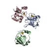
| ||||||||
|---|---|---|---|---|---|---|---|---|---|
| 1 | 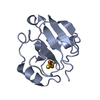
| ||||||||
| 2 | 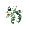
| ||||||||
| 3 | 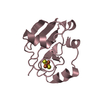
| ||||||||
| 4 | 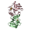
| ||||||||
| Unit cell |
|
-Components
| #1: Protein | Mass: 11412.846 Da / Num. of mol.: 3 / Mutation: C73S Source method: isolated from a genetically manipulated source Source: (gene. exp.) Pseudomonas putida (bacteria) / Gene: CAMB / Plasmid: pET / Species (production host): Escherichia coli / Production host: Escherichia coli BL21(DE3) (bacteria) / Strain (production host): BL21(DE3) / References: UniProt: P00259#2: Chemical | Iron–sulfur cluster  Mass: 175.820 Da / Num. of mol.: 3 / Source method: obtained synthetically / Formula: Fe2S2 Mass: 175.820 Da / Num. of mol.: 3 / Source method: obtained synthetically / Formula: Fe2S2#3: Water | ChemComp-HOH / | Water Mass: 18.015 Da / Num. of mol.: 224 / Source method: isolated from a natural source / Formula: H2O Mass: 18.015 Da / Num. of mol.: 224 / Source method: isolated from a natural source / Formula: H2O |
|---|
-Experimental details
-Experiment
| Experiment | Method: X-RAY DIFFRACTION / Number of used crystals: 1 |
|---|
- Sample preparation
| Crystal | Density Matthews: 2.28 Å3/Da / Density % sol: 45.53 % | ||||||||||||||||||||||||||||||
|---|---|---|---|---|---|---|---|---|---|---|---|---|---|---|---|---|---|---|---|---|---|---|---|---|---|---|---|---|---|---|---|
| Crystal grow | Temperature: 298 K / Method: vapor diffusion, hanging drop / pH: 5.9 Details: lithium sulfate, sodium citrate, dithiothreitol, pH 5.9, VAPOR DIFFUSION, HANGING DROP, temperature 298K | ||||||||||||||||||||||||||||||
| Crystal grow | *PLUS pH: 7.4 / Method: vapor diffusion, hanging drop | ||||||||||||||||||||||||||||||
| Components of the solutions | *PLUS
|
-Data collection
| Diffraction | Mean temperature: 110 K |
|---|---|
| Diffraction source | Source: SYNCHROTRON / Site: SSRL  / Beamline: BL7-1 / Wavelength: 1.08 Å / Beamline: BL7-1 / Wavelength: 1.08 Å |
| Detector | Type: MARRESEARCH / Detector: IMAGE PLATE / Date: Dec 18, 2002 / Details: mirrors |
| Radiation | Monochromator: Yale mirrors / Protocol: SINGLE WAVELENGTH / Monochromatic (M) / Laue (L): M / Scattering type: x-ray |
| Radiation wavelength | Wavelength: 1.08 Å / Relative weight: 1 |
| Reflection | Resolution: 1.65→50 Å / Num. all: 181573 / Num. obs: 180222 / % possible obs: 96 % / Observed criterion σ(F): 1 / Observed criterion σ(I): -3 / Redundancy: 4.5 % / Biso Wilson estimate: 19.9 Å2 / Rmerge(I) obs: 0.093 / Rsym value: 0.093 / Net I/σ(I): 6.5 |
| Reflection shell | Resolution: 1.65→1.68 Å / Redundancy: 4 % / Rmerge(I) obs: 0.618 / Mean I/σ(I) obs: 1.88 / Num. unique all: 1996 / Rsym value: 0.618 / % possible all: 97.8 |
| Reflection | *PLUS Highest resolution: 1.67 Å / Num. obs: 39004 / % possible obs: 96.7 % / Rmerge(I) obs: 0.055 |
- Processing
| Software |
| |||||||||||||||||||||||||
|---|---|---|---|---|---|---|---|---|---|---|---|---|---|---|---|---|---|---|---|---|---|---|---|---|---|---|
| Refinement | Method to determine structure: MOLECULAR REPLACEMENT Starting model: putidaredoxin C73S/C85S mutant, PDB ID code 1OQQ Resolution: 1.65→27.65 Å / Rfactor Rfree error: 0.006 / Isotropic thermal model: RESTRAINED / Cross valid method: THROUGHOUT / σ(F): 0 / σ(I): -3
| |||||||||||||||||||||||||
| Solvent computation | Solvent model: FLAT MODEL / Bsol: 34.0092 Å2 / ksol: 0.400588 e/Å3 | |||||||||||||||||||||||||
| Displacement parameters | Biso mean: 22.7 Å2
| |||||||||||||||||||||||||
| Refine analyze |
| |||||||||||||||||||||||||
| Refinement step | Cycle: LAST / Resolution: 1.65→27.65 Å
| |||||||||||||||||||||||||
| Refine LS restraints |
| |||||||||||||||||||||||||
| LS refinement shell | Resolution: 1.65→1.71 Å / Rfactor Rfree error: 0.024 / Total num. of bins used: 10
| |||||||||||||||||||||||||
| Xplor file |
| |||||||||||||||||||||||||
| Refinement | *PLUS Highest resolution: 1.67 Å / % reflection Rfree: 5 % | |||||||||||||||||||||||||
| Solvent computation | *PLUS | |||||||||||||||||||||||||
| Displacement parameters | *PLUS | |||||||||||||||||||||||||
| Refine LS restraints | *PLUS
|
