 Movie
Movie Controller

[English] 日本語
 Yorodumi
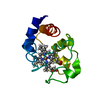
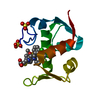
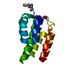
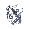
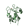
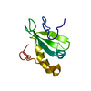
- PDB-2cxd: Crystal structure of conserved hypothetical protein, TTHA0068 fro... -
+ Open data

- Basic information
| Entry | Database: PDB / ID: 2cxd | ||||||
|---|---|---|---|---|---|---|---|
| Title | Crystal structure of conserved hypothetical protein, TTHA0068 from Thermus thermophilus HB8 | ||||||
 Components Components | conserved hypothetical protein, TTHA0068 | ||||||
 Keywords Keywords |  STRUCTURAL GENOMICS / UNKNOWN FUNCTION / conserved hypothetical protein / NPPSFA / National Project on Protein Structural and Functional Analyses / RIKEN Structural Genomics/Proteomics Initiative / RSGI STRUCTURAL GENOMICS / UNKNOWN FUNCTION / conserved hypothetical protein / NPPSFA / National Project on Protein Structural and Functional Analyses / RIKEN Structural Genomics/Proteomics Initiative / RSGI | ||||||
| Function / homology | TTHA0068-like / Protein of unknown function DUF309 / TTHA0068-like superfamily / Domain of unknown function (DUF309) / Hyaluronidase domain-like / Orthogonal Bundle / Mainly Alpha / DUF309 domain-containing protein Function and homology information Function and homology information | ||||||
| Biological species |   Thermus thermophilus (bacteria) Thermus thermophilus (bacteria) | ||||||
| Method | X-RAY DIFFRACTION / SYNCHROTRON / MOLECULAR REPLACEMENT / Resolution: 2 Å | ||||||
 Authors Authors | Kishishita, S. / Murayama, K. / Shirouzu, M. / Yokoyama, S. / RIKEN Structural Genomics/Proteomics Initiative (RSGI) | ||||||
 Citation Citation | Journal: To be Published Title: Crystal structure of conserved hypothetical protein, TTHA0068 from Thermus thermophilus HB8 Authors: Kishishita, S. / Murayama, K. / Shirouzu, M. / Yokoyama, S. | ||||||
| History |
|
- Structure visualization
| Structure viewer | Molecule: MolmilJmol/JSmol |
|---|
- Downloads & links
-Download
| PDBx/mmCIF format | 2cxd.cif.gz | 53.4 KB | Display | PDBx/mmCIF format |
|---|---|---|---|---|
| PDB format | pdb2cxd.ent.gz | 38.5 KB | Display | PDB format |
| PDBx/mmJSON format | 2cxd.json.gz | Tree view | PDBx/mmJSON format | |
| Others |  Other downloads Other downloads |
-Validation report
| Arichive directory | https://data.pdbj.org/pub/pdb/validation_reports/cx/2cxdftp://data.pdbj.org/pub/pdb/validation_reports/cx/2cxd | HTTPS FTP |
|---|
-Related structure data
| Related structure data |  2cwySC S: Starting model for refinement C: citing same article ( |
|---|---|
| Similar structure data | |
| Other databases |

-Links
 PDBj
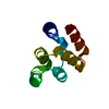
PDBj- Assembly
| Deposited unit | 
| ||||||||
|---|---|---|---|---|---|---|---|---|---|
| 1 | 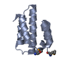
| ||||||||
| 2 | 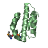
| ||||||||
| Unit cell |
| ||||||||
| Details | In this crystal packing, it looks like monomer. But, there is no experimental evidence. |
-Components
| #1: Protein | Mass: 11052.597 Da / Num. of mol.: 2 Source method: isolated from a genetically manipulated source Source: (gene. exp.) Thermus thermophilus (bacteria) / Strain: HB8 / Plasmid: pET11 / Production host: Escherichia coli (E. coli) / Strain (production host): B834(DE3) / References: UniProt: Q5SM75#2: Water | ChemComp-HOH / | Water Mass: 18.015 Da / Num. of mol.: 186 / Source method: isolated from a natural source / Formula: H2O Mass: 18.015 Da / Num. of mol.: 186 / Source method: isolated from a natural source / Formula: H2O |
|---|
-Experimental details
-Experiment
| Experiment | Method: X-RAY DIFFRACTION / Number of used crystals: 1 |
|---|
- Sample preparation
| Crystal | Density Matthews: 1.9 Å3/Da / Density % sol: 36 % |
|---|---|
| Crystal grow | Temperature: 293 K / Method: vapor diffusion, sitting drop / pH: 5.5 Details: 0.1M Bis-Tris, 25%(w/v) PEG 3350, pH 5.5, VAPOR DIFFUSION, SITTING DROP, temperature 293K |
-Data collection
| Diffraction | Mean temperature: 100 K |
|---|---|
| Diffraction source | Source: SYNCHROTRON / Site: SPring-8  / Beamline: BL26B1 / Wavelength: 0.964 Å / Beamline: BL26B1 / Wavelength: 0.964 Å |
| Detector | Type: RIGAKU JUPITER 210 / Detector: CCD / Date: May 27, 2005 / Details: mirrors |
| Radiation | Monochromator: silicon / Protocol: SINGLE WAVELENGTH / Monochromatic (M) / Laue (L): M / Scattering type: x-ray |
| Radiation wavelength | Wavelength: 0.964 Å / Relative weight: 1 |
| Reflection | Resolution: 2→50 Å / Num. obs: 12010 / % possible obs: 99.9 % / Observed criterion σ(I): -3 / Redundancy: 6.9 % / Biso Wilson estimate: 10.4 Å2 / Rsym value: 0.084 / Net I/σ(I): 20.3 |
| Reflection shell | Resolution: 2→2.09 Å / Mean I/σ(I) obs: 8 / Rsym value: 0.275 / % possible all: 99.9 |
- Processing
| Software |
| ||||||||||||||||||||||||||||||||||||||||||||||||||||||||||||
|---|---|---|---|---|---|---|---|---|---|---|---|---|---|---|---|---|---|---|---|---|---|---|---|---|---|---|---|---|---|---|---|---|---|---|---|---|---|---|---|---|---|---|---|---|---|---|---|---|---|---|---|---|---|---|---|---|---|---|---|---|---|
| Refinement | Method to determine structure: MOLECULAR REPLACEMENT Starting model: PDB ENTRY 2CWY Resolution: 2→39.6 Å / Rfactor Rfree error: 0.009 / Data cutoff high absF: 1225920.95 / Data cutoff low absF: 0 / Isotropic thermal model: RESTRAINED / Cross valid method: THROUGHOUT / σ(F): 0
| ||||||||||||||||||||||||||||||||||||||||||||||||||||||||||||
| Solvent computation | Solvent model: FLAT MODEL / Bsol: 35.1658 Å2 / ksol: 0.315936 e/Å3 | ||||||||||||||||||||||||||||||||||||||||||||||||||||||||||||
| Displacement parameters | Biso mean: 21.2 Å2
| ||||||||||||||||||||||||||||||||||||||||||||||||||||||||||||
| Refine analyze |
| ||||||||||||||||||||||||||||||||||||||||||||||||||||||||||||
| Refinement step | Cycle: LAST / Resolution: 2→39.6 Å
| ||||||||||||||||||||||||||||||||||||||||||||||||||||||||||||
| Refine LS restraints |
| ||||||||||||||||||||||||||||||||||||||||||||||||||||||||||||
| LS refinement shell | Resolution: 2→2.13 Å / Rfactor Rfree error: 0.02 / Total num. of bins used: 6
| ||||||||||||||||||||||||||||||||||||||||||||||||||||||||||||
| Xplor file |
|
