 Movie
Movie Controller

[English] 日本語
 Yorodumi
- PDB-1on0: Crystal Structure of Putative Acetyltransferase (YycN) from Bacil... -
+ Open data

- Basic information
| Entry | Database: PDB / ID: 1on0 | ||||||
|---|---|---|---|---|---|---|---|
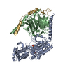
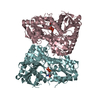
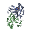
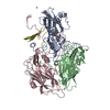
| Title | Crystal Structure of Putative Acetyltransferase (YycN) from Bacillus subtilis, NORTHEAST STRUCTURAL GENOMICS CONSORTIUM TARGET SR144 | ||||||
 Components Components | YycN protein | ||||||
 Keywords Keywords |  STRUCTURAL GENOMICS / UNKNOWN FUNCTION / alpha-beta protein with anti-parallel beta strands / PSI / Protein Structure Initiative / Northeast Structural Genomics Consortium / NESG STRUCTURAL GENOMICS / UNKNOWN FUNCTION / alpha-beta protein with anti-parallel beta strands / PSI / Protein Structure Initiative / Northeast Structural Genomics Consortium / NESG | ||||||
| Function / homology |  Function and homology information Function and homology informationacyltransferase activity, transferring groups other than amino-acyl groups / Transferases; Acyltransferases; Transferring groups other than aminoacyl groupsSimilarity search - Function | ||||||
| Biological species |  Bacillus subtilis (bacteria) Bacillus subtilis (bacteria) | ||||||
| Method | X-RAY DIFFRACTION / SYNCHROTRON / SAD / Resolution: 2.2 Å | ||||||
 Authors Authors | Forouhar, F. / Shen, J. / Kuzin, A. / Chiang, Y. / Xiao, R. / Acton, T.B. / Rost, B. / Montelione, G.T. / Tong, L. / Northeast Structural Genomics Consortium (NESG) | ||||||
 Citation Citation | Journal: To be Published Title: Crystal Structure of Putative Acetyltransferase (YycN) from Bacillus subtilis Authors: Forouhar, F. / Shen, J. / Kuzin, A. / Chiang, Y. / Xiao, R. / Acton, T.B. / Rost, B. / Montelione, G.T. / Tong, L. | ||||||
| History |
|
- Structure visualization
| Structure viewer | Molecule: MolmilJmol/JSmol |
|---|
- Downloads & links
-Download
| PDBx/mmCIF format | 1on0.cif.gz | 141.2 KB | Display | PDBx/mmCIF format |
|---|---|---|---|---|
| PDB format | pdb1on0.ent.gz | 118.5 KB | Display | PDB format |
| PDBx/mmJSON format | 1on0.json.gz | Tree view | PDBx/mmJSON format | |
| Others |  Other downloads Other downloads |
-Validation report
| Arichive directory | https://data.pdbj.org/pub/pdb/validation_reports/on/1on0ftp://data.pdbj.org/pub/pdb/validation_reports/on/1on0 | HTTPS FTP |
|---|
-Related structure data
| Similar structure data | |
|---|---|
| Other databases |


-Links
 PDBj
PDBj

- Assembly
| Deposited unit | 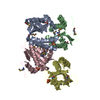
| ||||||||
|---|---|---|---|---|---|---|---|---|---|
| 1 |
| ||||||||
| 2 | 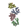
| ||||||||
| Unit cell |
|
-Components
| #1: Protein | Mass: 18667.529 Da / Num. of mol.: 4 Source method: isolated from a genetically manipulated source Source: (gene. exp.) Bacillus subtilis (bacteria) / Gene: YYCN / Plasmid: pET21 / Production host: Escherichia coli (E. coli) / Strain (production host): BL21 / References: UniProt: O32293#2: Chemical | ChemComp-SO4 / Sulfate  Mass: 96.063 Da / Num. of mol.: 4 / Source method: obtained synthetically / Formula: SO4 Mass: 96.063 Da / Num. of mol.: 4 / Source method: obtained synthetically / Formula: SO4#3: Chemical | ChemComp-CL / | Chloride  Mass: 35.453 Da / Num. of mol.: 1 / Source method: obtained synthetically / Formula: Cl Mass: 35.453 Da / Num. of mol.: 1 / Source method: obtained synthetically / Formula: Cl#4: Water | ChemComp-HOH / | Water Mass: 18.015 Da / Num. of mol.: 357 / Source method: isolated from a natural source / Formula: H2O Mass: 18.015 Da / Num. of mol.: 357 / Source method: isolated from a natural source / Formula: H2O |
|---|
-Experimental details
-Experiment
| Experiment | Method: X-RAY DIFFRACTION / Number of used crystals: 1 |
|---|
- Sample preparation
| Crystal | Density Matthews: 2.5 Å3/Da / Density % sol: 50.51 % |
|---|---|
| Crystal grow | Temperature: 293 K / Method: vapor diffusion, hanging drop / pH: 7.5 Details: 5mM Tris, 20% PEG 8k, 0.2 M Ammonium Sulfate, 50 mM Sodium Chloride, and 5 mM DTT, pH 7.5, VAPOR DIFFUSION, HANGING DROP, temperature 293K |
-Data collection
| Diffraction | Mean temperature: 100 K |
|---|---|
| Diffraction source | Source: SYNCHROTRON / Site: NSLS  / Beamline: X4A / Wavelength: 0.982 Å / Beamline: X4A / Wavelength: 0.982 Å |
| Detector | Type: ADSC QUANTUM 4 / Detector: CCD / Date: Feb 3, 2003 |
| Radiation | Protocol: SINGLE WAVELENGTH / Monochromatic (M) / Laue (L): M / Scattering type: x-ray |
| Radiation wavelength | Wavelength: 0.982 Å / Relative weight: 1 |
| Reflection | Resolution: 2.2→29.68 Å / Num. all: 76470 / Num. obs: 69770 / % possible obs: 91.5 % / Observed criterion σ(F): -3 / Observed criterion σ(I): -3 / Redundancy: 3.26 % / Biso Wilson estimate: 16.7 Å2 / Rmerge(I) obs: 0.071 / Rsym value: 0.069 / Net I/σ(I): 16.64 |
| Reflection shell | Resolution: 2.2→2.28 Å / Redundancy: 2.7 % / Rmerge(I) obs: 0.354 / Mean I/σ(I) obs: 3.74 / Num. unique all: 2778 / Rsym value: 0.287 / % possible all: 99.1 |
- Processing
| Software |
| |||||||||||||||||||||||||
|---|---|---|---|---|---|---|---|---|---|---|---|---|---|---|---|---|---|---|---|---|---|---|---|---|---|---|
| Refinement | Method to determine structure: SAD / Resolution: 2.2→29.68 Å / Cross valid method: THROUGHOUT / σ(F): 2
| |||||||||||||||||||||||||
| Displacement parameters | Biso mean: 35.8 Å2
| |||||||||||||||||||||||||
| Refine analyze |
| |||||||||||||||||||||||||
| Refinement step | Cycle: LAST / Resolution: 2.2→29.68 Å
| |||||||||||||||||||||||||
| Refine LS restraints |
| |||||||||||||||||||||||||
| LS refinement shell | Resolution: 2.2→29.68 Å / Rfactor Rfree error: 0.003
|
