 Movie
Movie Controller

[English] 日本語
 Yorodumi
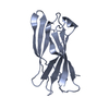
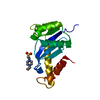
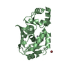
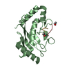
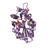
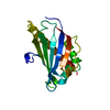
- PDB-1om9: Structure of the GGA1-appendage in complex with the p56 binding p... -
+ Open data

- Basic information
| Entry | Database: PDB / ID: 1om9 | ||||||
|---|---|---|---|---|---|---|---|
| Title | Structure of the GGA1-appendage in complex with the p56 binding peptide | ||||||
 Components Components |
| ||||||
 Keywords Keywords |  PROTEIN TRANSPORT / SIGNALING PROTEIN / beta sandwich / beta augmentation / GGA / adaptin / clathrin adaptor PROTEIN TRANSPORT / SIGNALING PROTEIN / beta sandwich / beta augmentation / GGA / adaptin / clathrin adaptor | ||||||
| Function / homology |  Function and homology information Function and homology informationGolgi to lysosome transport / protein localization to ciliary membrane / Golgi to plasma membrane protein transport / Golgi to plasma membrane transport / protein localization to cell surface / retrograde transport, endosome to Golgi / TBC/RABGAPs / phosphatidylinositol binding / ubiquitin binding / intracellular protein transport ...Golgi to lysosome transport / protein localization to ciliary membrane / Golgi to plasma membrane protein transport / Golgi to plasma membrane transport / protein localization to cell surface / retrograde transport, endosome to Golgi / TBC/RABGAPs / phosphatidylinositol binding / ubiquitin binding / intracellular protein transport / protein catabolic process / trans-Golgi network / protein localization / small GTPase binding / positive regulation of protein catabolic process / protein transport / early endosome membrane / early endosome / endosome membrane / Amyloid fiber formation / intracellular membrane-bounded organelle / Golgi apparatus / protein-containing complex / nucleoplasm / membrane / identical protein binding / cytosolSimilarity search - Function | ||||||
| Biological species |  Homo sapiens (human) Homo sapiens (human) | ||||||
| Method | X-RAY DIFFRACTION / MOLECULAR REPLACEMENT / Resolution: 2.5 Å | ||||||
 Authors Authors | Collins, B.M. / Praefcke, G.J.K. / Robinson, M.S. / Owen, D.J. | ||||||
 Citation Citation | Journal: Nat.Struct.Biol. / Year: 2003 Title: Structural basis for binding of accessory proteins by the appendage domain of GGAs Authors: Collins, B.M. / Praefcke, G.J.K. / Robinson, M.S. / Owen, D.J. | ||||||
| History |
|
- Structure visualization
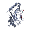
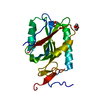
| Structure viewer | Molecule: MolmilJmol/JSmol |
|---|
- Downloads & links
-Download
| PDBx/mmCIF format | 1om9.cif.gz | 72.6 KB | Display | PDBx/mmCIF format |
|---|---|---|---|---|
| PDB format | pdb1om9.ent.gz | 54.7 KB | Display | PDB format |
| PDBx/mmJSON format | 1om9.json.gz | Tree view | PDBx/mmJSON format | |
| Others |  Other downloads Other downloads |
-Validation report
| Arichive directory | https://data.pdbj.org/pub/pdb/validation_reports/om/1om9ftp://data.pdbj.org/pub/pdb/validation_reports/om/1om9 | HTTPS FTP |
|---|
-Related structure data
| Related structure data |  1na8S S: Starting model for refinement |
|---|---|
| Similar structure data |


-Links
 PDBj
PDBj


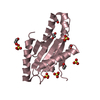
- Assembly
| Deposited unit | 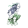
| ||||||||||||||||||||||||||||||||||||||||||||||||||||||||||||||||||||
|---|---|---|---|---|---|---|---|---|---|---|---|---|---|---|---|---|---|---|---|---|---|---|---|---|---|---|---|---|---|---|---|---|---|---|---|---|---|---|---|---|---|---|---|---|---|---|---|---|---|---|---|---|---|---|---|---|---|---|---|---|---|---|---|---|---|---|---|---|---|
| 1 | 
| ||||||||||||||||||||||||||||||||||||||||||||||||||||||||||||||||||||
| 2 | 
| ||||||||||||||||||||||||||||||||||||||||||||||||||||||||||||||||||||
| 3 |
| ||||||||||||||||||||||||||||||||||||||||||||||||||||||||||||||||||||
| Unit cell |
| ||||||||||||||||||||||||||||||||||||||||||||||||||||||||||||||||||||
| Noncrystallographic symmetry (NCS) | NCS domain:
NCS domain segments: Component-ID: 1 / Refine code: 2
NCS ensembles :
| ||||||||||||||||||||||||||||||||||||||||||||||||||||||||||||||||||||
| Details | Protein chain A is paired with peptide chain P / Protein chain B is paired with peptide chain Q |
-Components
| #1: Protein | Mass: 17323.117 Da / Num. of mol.: 2 / Fragment: appendage domain, Residues 494-639 of SWS Q9UJY5 Source method: isolated from a genetically manipulated source Source: (gene. exp.) Homo sapiens (human) / Gene: GGA1 / Plasmid: pMWH6172 / Production host:  Escherichia coli (E. coli) / Strain (production host): BL21(DE3)/pLysS / References: UniProt: Q9UJY5 Escherichia coli (E. coli) / Strain (production host): BL21(DE3)/pLysS / References: UniProt: Q9UJY5#2: Protein/peptide | Mass: 1650.566 Da / Num. of mol.: 2 / Source method: obtained synthetically / Details: sequence occurs naturally in Homo sapiens / References: UniProt: Q7Z6B0*PLUS #3: Water | ChemComp-HOH / | Water Mass: 18.015 Da / Num. of mol.: 66 / Source method: isolated from a natural source / Formula: H2O Mass: 18.015 Da / Num. of mol.: 66 / Source method: isolated from a natural source / Formula: H2O |
|---|
-Experimental details
-Experiment
| Experiment | Method: X-RAY DIFFRACTION / Number of used crystals: 1 |
|---|
- Sample preparation
| Crystal | Density Matthews: 2.31 Å3/Da / Density % sol: 46.2 % | ||||||||||||||||||||||||||||||||||||||||||
|---|---|---|---|---|---|---|---|---|---|---|---|---|---|---|---|---|---|---|---|---|---|---|---|---|---|---|---|---|---|---|---|---|---|---|---|---|---|---|---|---|---|---|---|
| Crystal grow | Temperature: 288 K / Method: vapor diffusion, sitting drop / pH: 5.5 Details: 100 mM sodium citrate, 100 mM MgCl2 and 35% PEG 400, pH 5.5, VAPOR DIFFUSION, SITTING DROP, temperature 288K | ||||||||||||||||||||||||||||||||||||||||||
| Crystal grow | *PLUS pH: 7.5 / Method: vapor diffusion, sitting drop | ||||||||||||||||||||||||||||||||||||||||||
| Components of the solutions | *PLUS
|
-Data collection
| Diffraction | Mean temperature: 100 K |
|---|---|
| Diffraction source | Source: ROTATING ANODE / Type: RIGAKU / Wavelength: 1.5418 Å |
| Detector | Type: MARRESEARCH / Detector: IMAGE PLATE / Date: Jan 5, 2003 |
| Radiation | Protocol: SINGLE WAVELENGTH / Monochromatic (M) / Laue (L): M / Scattering type: x-ray |
| Radiation wavelength | Wavelength: 1.5418 Å / Relative weight: 1 |
| Reflection | Resolution: 2.5→30 Å / Num. all: 11523 / Num. obs: 11512 / % possible obs: 99.9 % / Observed criterion σ(I): 3.1 / Redundancy: 4.9 % / Biso Wilson estimate: 55.4 Å2 / Rmerge(I) obs: 0.081 / Net I/σ(I): 16.5 |
| Reflection shell | Resolution: 2.5→2.64 Å / Redundancy: 5 % / Rmerge(I) obs: 0.478 / Mean I/σ(I) obs: 3.1 / % possible all: 99.9 |
| Reflection | *PLUS Highest resolution: 2.5 Å / Lowest resolution: 30 Å |
| Reflection shell | *PLUS % possible obs: 99.9 % |
- Processing
| Software |
| ||||||||||||||||||||||||||||||||||||||||||||||||||||||||||||||||||||||||||||||||||||||||||||||||||||||||||||||||||||||||||||||||||
|---|---|---|---|---|---|---|---|---|---|---|---|---|---|---|---|---|---|---|---|---|---|---|---|---|---|---|---|---|---|---|---|---|---|---|---|---|---|---|---|---|---|---|---|---|---|---|---|---|---|---|---|---|---|---|---|---|---|---|---|---|---|---|---|---|---|---|---|---|---|---|---|---|---|---|---|---|---|---|---|---|---|---|---|---|---|---|---|---|---|---|---|---|---|---|---|---|---|---|---|---|---|---|---|---|---|---|---|---|---|---|---|---|---|---|---|---|---|---|---|---|---|---|---|---|---|---|---|---|---|---|---|
| Refinement | Method to determine structure: MOLECULAR REPLACEMENT Starting model: PDB ID 1NA8 chain A Resolution: 2.5→30 Å / Cor.coef. Fo:Fc: 0.936 / Cor.coef. Fo:Fc free: 0.916 / SU B: 10.434 / SU ML: 0.231 / Cross valid method: THROUGHOUT / σ(F): 1.55 / σ(I): 3.1 / ESU R: 0.784 / ESU R Free: 0.315 / Stereochemistry target values: MAXIMUM LIKELIHOOD / Details: HYDROGENS HAVE BEEN ADDED IN THE RIDING POSITIONS
| ||||||||||||||||||||||||||||||||||||||||||||||||||||||||||||||||||||||||||||||||||||||||||||||||||||||||||||||||||||||||||||||||||
| Solvent computation | Ion probe radii: 0.8 Å / Shrinkage radii: 0.8 Å / VDW probe radii: 1.4 Å / Solvent model: BABINET MODEL WITH MASK | ||||||||||||||||||||||||||||||||||||||||||||||||||||||||||||||||||||||||||||||||||||||||||||||||||||||||||||||||||||||||||||||||||
| Displacement parameters | Biso mean: 42.167 Å2
| ||||||||||||||||||||||||||||||||||||||||||||||||||||||||||||||||||||||||||||||||||||||||||||||||||||||||||||||||||||||||||||||||||
| Refinement step | Cycle: LAST / Resolution: 2.5→30 Å
| ||||||||||||||||||||||||||||||||||||||||||||||||||||||||||||||||||||||||||||||||||||||||||||||||||||||||||||||||||||||||||||||||||
| Refine LS restraints |
| ||||||||||||||||||||||||||||||||||||||||||||||||||||||||||||||||||||||||||||||||||||||||||||||||||||||||||||||||||||||||||||||||||
| Refine LS restraints NCS | Refine-ID: X-RAY DIFFRACTION
| ||||||||||||||||||||||||||||||||||||||||||||||||||||||||||||||||||||||||||||||||||||||||||||||||||||||||||||||||||||||||||||||||||
| LS refinement shell | Resolution: 2.5→2.565 Å / Total num. of bins used: 20 /
| ||||||||||||||||||||||||||||||||||||||||||||||||||||||||||||||||||||||||||||||||||||||||||||||||||||||||||||||||||||||||||||||||||
| Refinement | *PLUS Highest resolution: 2.5 Å / Rfactor Rfree: 0.262 / Rfactor Rwork: 0.216 | ||||||||||||||||||||||||||||||||||||||||||||||||||||||||||||||||||||||||||||||||||||||||||||||||||||||||||||||||||||||||||||||||||
| Solvent computation | *PLUS | ||||||||||||||||||||||||||||||||||||||||||||||||||||||||||||||||||||||||||||||||||||||||||||||||||||||||||||||||||||||||||||||||||
| Displacement parameters | *PLUS | ||||||||||||||||||||||||||||||||||||||||||||||||||||||||||||||||||||||||||||||||||||||||||||||||||||||||||||||||||||||||||||||||||
| Refine LS restraints | *PLUS
| ||||||||||||||||||||||||||||||||||||||||||||||||||||||||||||||||||||||||||||||||||||||||||||||||||||||||||||||||||||||||||||||||||
| LS refinement shell | *PLUS Highest resolution: 2.5 Å / Lowest resolution: 2.56 Å |
