 Movie
Movie Controller

[English] 日本語
 Yorodumi
- PDB-6eib: Structure of the active GGEEF domain of a diguanylate cyclase fro... -
+ Open data

- Basic information
| Entry | Database: PDB / ID: 6eib | ||||||
|---|---|---|---|---|---|---|---|
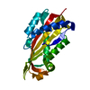
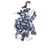
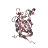
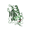
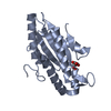
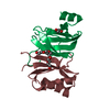
| Title | Structure of the active GGEEF domain of a diguanylate cyclase from Vibrio cholerae. | ||||||
 Components Components | Sensory box/GGDEF family protein | ||||||
 Keywords Keywords |  TRANSFERASE / diguanylate cyclase / GGEEF domain TRANSFERASE / diguanylate cyclase / GGEEF domain | ||||||
| Function / homology |  Function and homology informationdiguanylate cyclase / diguanylate cyclase activity / GTP binding / metal ion binding Function and homology informationdiguanylate cyclase / diguanylate cyclase activity / GTP binding / metal ion bindingSimilarity search - Function | ||||||
| Biological species |  Vibrio cholerae serotype O1 (bacteria) Vibrio cholerae serotype O1 (bacteria) | ||||||
| Method | X-RAY DIFFRACTION / SYNCHROTRON / MOLECULAR REPLACEMENT / Resolution: 1.941 Å | ||||||
 Authors Authors | Chouhan, O.P. / Roske, Y. | ||||||
| Funding support |  Germany, 1items Germany, 1items
| ||||||
 Citation Citation | Journal: Biochem.Biophys.Res.Commun. / Year: 2020 Title: Structure of the active GGEEF domain of a diguanylate cyclase from Vibrio cholerae. Authors: Chouhan, O.P. / Roske, Y. / Heinemann, U. / Biswas, S. | ||||||
| History |
|
- Structure visualization
| Structure viewer | Molecule: MolmilJmol/JSmol |
|---|
- Downloads & links
-Download
| PDBx/mmCIF format | 6eib.cif.gz | 288.4 KB | Display | PDBx/mmCIF format |
|---|---|---|---|---|
| PDB format | pdb6eib.ent.gz | 235.7 KB | Display | PDB format |
| PDBx/mmJSON format | 6eib.json.gz | Tree view | PDBx/mmJSON format | |
| Others |  Other downloads Other downloads |
-Validation report
| Arichive directory | https://data.pdbj.org/pub/pdb/validation_reports/ei/6eibftp://data.pdbj.org/pub/pdb/validation_reports/ei/6eib | HTTPS FTP |
|---|
-Related structure data
| Related structure data | 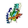 4zveS S: Starting model for refinement |
|---|---|
| Similar structure data |


-Links
 PDBj
PDBj


- Assembly
| Deposited unit | 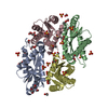
| ||||||||
|---|---|---|---|---|---|---|---|---|---|
| 1 | 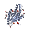
| ||||||||
| 2 | 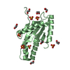
| ||||||||
| 3 | 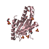
| ||||||||
| 4 | 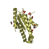
| ||||||||
| Unit cell |
|
-Components
| #1: Protein | Mass: 18951.459 Da / Num. of mol.: 4 Source method: isolated from a genetically manipulated source Source: (gene. exp.) Vibrio cholerae serotype O1 (strain ATCC 39541 / Classical Ogawa 395 / O395) (bacteria)Gene: VC0395_0300 / Plasmid: pGEX_6P1 / Production host: Escherichia coli (E. coli) / Strain (production host): BL21 / References: UniProt: A0A0H3AFM6#2: Chemical | ChemComp-SO4 / Sulfate  Mass: 96.063 Da / Num. of mol.: 18 / Source method: obtained synthetically / Formula: SO4 Mass: 96.063 Da / Num. of mol.: 18 / Source method: obtained synthetically / Formula: SO4#3: Chemical | ChemComp-EDO / Ethylene glycol  Mass: 62.068 Da / Num. of mol.: 22 / Source method: obtained synthetically / Formula: C2H6O2 Mass: 62.068 Da / Num. of mol.: 22 / Source method: obtained synthetically / Formula: C2H6O2#4: Chemical | Acetate  Mass: 59.044 Da / Num. of mol.: 3 / Source method: obtained synthetically / Formula: C2H3O2 Mass: 59.044 Da / Num. of mol.: 3 / Source method: obtained synthetically / Formula: C2H3O2#5: Water | ChemComp-HOH / | Water Mass: 18.015 Da / Num. of mol.: 521 / Source method: isolated from a natural source / Formula: H2O Mass: 18.015 Da / Num. of mol.: 521 / Source method: isolated from a natural source / Formula: H2O |
|---|
-Experimental details
-Experiment
| Experiment | Method: X-RAY DIFFRACTION / Number of used crystals: 1 |
|---|
- Sample preparation
| Crystal | Density Matthews: 2.51 Å3/Da / Density % sol: 51.01 % |
|---|---|
| Crystal grow | Temperature: 277 K / Method: evaporation / pH: 5.2 / Details: 0.5M Ammonium sulfate, 0.1M Sodium citrate |
-Data collection
| Diffraction | Mean temperature: 100 K |
|---|---|
| Diffraction source | Source: SYNCHROTRON / Site: BESSY / Beamline: 14.1 / Wavelength: 0.98141 Å |
| Detector | Type: DECTRIS PILATUS3 6M / Detector: PIXEL / Date: Mar 5, 2017 |
| Radiation | Protocol: SINGLE WAVELENGTH / Monochromatic (M) / Laue (L): M / Scattering type: x-ray |
| Radiation wavelength | Wavelength: 0.98141 Å / Relative weight: 1 |
| Reflection | Resolution: 1.94→43.41 Å / Num. obs: 52783 / % possible obs: 96.3 % / Redundancy: 2.34 % / Biso Wilson estimate: 32.69 Å2 / CC1/2: 0.995 / Rsym value: 0.094 / Net I/σ(I): 7.82 |
| Reflection shell | Resolution: 1.94→2.06 Å / Mean I/σ(I) obs: 1.26 / Num. unique obs: 8471 / CC1/2: 0.687 / Rsym value: 0.763 / % possible all: 95.9 |
- Processing
| Software |
| |||||||||||||||||||||||||||||||||||||||||||||||||||||||||||||||||||||||||||||||||||||||||||||||||||||||||||||||||||||||||||||
|---|---|---|---|---|---|---|---|---|---|---|---|---|---|---|---|---|---|---|---|---|---|---|---|---|---|---|---|---|---|---|---|---|---|---|---|---|---|---|---|---|---|---|---|---|---|---|---|---|---|---|---|---|---|---|---|---|---|---|---|---|---|---|---|---|---|---|---|---|---|---|---|---|---|---|---|---|---|---|---|---|---|---|---|---|---|---|---|---|---|---|---|---|---|---|---|---|---|---|---|---|---|---|---|---|---|---|---|---|---|---|---|---|---|---|---|---|---|---|---|---|---|---|---|---|---|---|
| Refinement | Method to determine structure: MOLECULAR REPLACEMENT Starting model: 4ZVE Resolution: 1.941→35.534 Å / SU ML: 0.26 / Cross valid method: FREE R-VALUE / σ(F): 1.36 / Phase error: 27.01
| |||||||||||||||||||||||||||||||||||||||||||||||||||||||||||||||||||||||||||||||||||||||||||||||||||||||||||||||||||||||||||||
| Solvent computation | Shrinkage radii: 0.9 Å / VDW probe radii: 1.11 Å | |||||||||||||||||||||||||||||||||||||||||||||||||||||||||||||||||||||||||||||||||||||||||||||||||||||||||||||||||||||||||||||
| Refinement step | Cycle: LAST / Resolution: 1.941→35.534 Å
| |||||||||||||||||||||||||||||||||||||||||||||||||||||||||||||||||||||||||||||||||||||||||||||||||||||||||||||||||||||||||||||
| Refine LS restraints |
| |||||||||||||||||||||||||||||||||||||||||||||||||||||||||||||||||||||||||||||||||||||||||||||||||||||||||||||||||||||||||||||
| LS refinement shell |
| |||||||||||||||||||||||||||||||||||||||||||||||||||||||||||||||||||||||||||||||||||||||||||||||||||||||||||||||||||||||||||||
| Refinement TLS params. | Method: refined / Refine-ID: X-RAY DIFFRACTION
| |||||||||||||||||||||||||||||||||||||||||||||||||||||||||||||||||||||||||||||||||||||||||||||||||||||||||||||||||||||||||||||
| Refinement TLS group |
|
