 Movie
Movie Controller

[English] 日本語
 Yorodumi
- PDB-1o9h: rRNA methyltransferase aviRa from Streptomyces viridochromogenes ... -
+ Open data

- Basic information
| Entry | Database: PDB / ID: 1o9h | ||||||
|---|---|---|---|---|---|---|---|
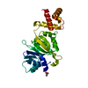
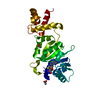
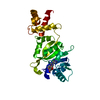
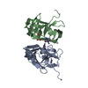
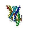
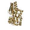
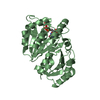
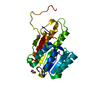
| Title | rRNA methyltransferase aviRa from Streptomyces viridochromogenes at 2.4A | ||||||
 Components Components | RRNA METHYLTRANSFERASE | ||||||
 Keywords Keywords |  TRANSFERASE / ANTIBIOTIC RESISTANCE / RRNA-METHYLTRANSFERASE TRANSFERASE / ANTIBIOTIC RESISTANCE / RRNA-METHYLTRANSFERASE | ||||||
| Function / homology |  Function and homology information23S rRNA (guanine2535-N1)-methyltransferase / rRNA (guanine-N1-)-methyltransferase activity / rRNA methylation / methylation / response to antibiotic Function and homology information23S rRNA (guanine2535-N1)-methyltransferase / rRNA (guanine-N1-)-methyltransferase activity / rRNA methylation / methylation / response to antibioticSimilarity search - Function | ||||||
| Biological species |  STREPTOMYCES VIRIDOCHROMOGENES (bacteria) STREPTOMYCES VIRIDOCHROMOGENES (bacteria) | ||||||
| Method | X-RAY DIFFRACTION / MOLECULAR REPLACEMENT / Resolution: 2.4 Å | ||||||
 Authors Authors | Mosbacher, T.G. / Schulz, G.E. | ||||||
 Citation Citation | Journal: J.Mol.Biol. / Year: 2003 Title: Crystal Structure of the Avilamycin Resistance-Conferring Methyltransferase Avira from Streptomyces Viridochromogenes Authors: Mosbacher, T.G. / Bechthold, A. / Schulz, G.E. | ||||||
| History |
|
- Structure visualization
| Structure viewer | Molecule: MolmilJmol/JSmol |
|---|
- Downloads & links
-Download
| PDBx/mmCIF format | 1o9h.cif.gz | 59.8 KB | Display | PDBx/mmCIF format |
|---|---|---|---|---|
| PDB format | pdb1o9h.ent.gz | 43.3 KB | Display | PDB format |
| PDBx/mmJSON format | 1o9h.json.gz | Tree view | PDBx/mmJSON format | |
| Others |  Other downloads Other downloads |
-Validation report
| Arichive directory | https://data.pdbj.org/pub/pdb/validation_reports/o9/1o9hftp://data.pdbj.org/pub/pdb/validation_reports/o9/1o9h | HTTPS FTP |
|---|
-Related structure data
| Related structure data |  1o9gSC S: Starting model for refinement C: citing same article ( |
|---|---|
| Similar structure data |


-Links
 PDBj
PDBj- Assembly
| Deposited unit | 
| ||||||||
|---|---|---|---|---|---|---|---|---|---|
| 1 |
| ||||||||
| Unit cell |
|
-Components
| #1: Protein | Mass: 26670.760 Da / Num. of mol.: 1 Source method: isolated from a genetically manipulated source Source: (gene. exp.) STREPTOMYCES VIRIDOCHROMOGENES (bacteria)Plasmid: PRSETB / Production host: ESCHERICHIA COLI (E. coli) / Strain (production host): BL21(DE3) / References: UniProt: Q9F5K5 |
|---|---|
| #2: Water | ChemComp-HOH / Water Mass: 18.015 Da / Num. of mol.: 29 / Source method: isolated from a natural source / Formula: H2O Mass: 18.015 Da / Num. of mol.: 29 / Source method: isolated from a natural source / Formula: H2O |
| Sequence details | SEQUENCE IN DATABASE INCORRECT FROM RESIDUE 180 TO 195. THE SWISS-PROT ACCESSION Q9F5K5 HAS THE ...SEQUENCE IN DATABASE INCORRECT FROM RESIDUE 180 TO 195. THE SWISS-PROT ACCESSION Q9F5K5 HAS THE SEQUENCE SARTGKGRCP |
-Experimental details
-Experiment
| Experiment | Method: X-RAY DIFFRACTION / Number of used crystals: 1 |
|---|
- Sample preparation
| Crystal | Density Matthews: 2.14 Å3/Da / Density % sol: 39 % | ||||||||||||||||||||||||
|---|---|---|---|---|---|---|---|---|---|---|---|---|---|---|---|---|---|---|---|---|---|---|---|---|---|
| Crystal grow | pH: 6.6 / Details: MES 6.6,PEG 20K 11%, pH 6.60 | ||||||||||||||||||||||||
| Crystal grow | *PLUS Temperature: 20 ℃ / Method: vapor diffusion, hanging drop / PH range low: 6.9 / PH range high: 6.6 | ||||||||||||||||||||||||
| Components of the solutions | *PLUS
|
-Data collection
| Diffraction | Mean temperature: 287 K |
|---|---|
| Diffraction source | Source: ROTATING ANODE / Type: RIGAKU RU200B / Wavelength: 1.5418 |
| Detector | Type: BRUKER / Detector: AREA DETECTOR / Date: Jan 11, 2002 |
| Radiation | Protocol: SINGLE WAVELENGTH / Monochromatic (M) / Laue (L): M / Scattering type: x-ray |
| Radiation wavelength | Wavelength: 1.5418 Å / Relative weight: 1 |
| Reflection | Resolution: 2.4→26.4 Å / Num. obs: 8039 / % possible obs: 90 % / Observed criterion σ(I): 2 / Redundancy: 2.3 % / Rmerge(I) obs: 0.071 / Net I/σ(I): 38.7 |
| Reflection shell | Resolution: 2.4→2.46 Å / Redundancy: 1.5 % / Rmerge(I) obs: 0.282 / Mean I/σ(I) obs: 3.2 / % possible all: 90 |
| Reflection | *PLUS Highest resolution: 2.4 Å / Lowest resolution: 30 Å / Num. obs: 8093 / % possible obs: 91 % / Redundancy: 2.3 % / Num. measured all: 19577 / Rmerge(I) obs: 0.057 |
| Reflection shell | *PLUS % possible obs: 91 % / Redundancy: 1.5 % / Num. unique obs: 921 / Rmerge(I) obs: 0.21 / Mean I/σ(I) obs: 3.2 |
- Processing
| Software |
| ||||||||||||||||||||||||||||||||||||||||||||||||||||||||||||||||||||||||||||||||||||||||||||||||||||||||||||||||||||||||||||||||||||||||||||||||||||||||||||||||||||||||||||||||||||||
|---|---|---|---|---|---|---|---|---|---|---|---|---|---|---|---|---|---|---|---|---|---|---|---|---|---|---|---|---|---|---|---|---|---|---|---|---|---|---|---|---|---|---|---|---|---|---|---|---|---|---|---|---|---|---|---|---|---|---|---|---|---|---|---|---|---|---|---|---|---|---|---|---|---|---|---|---|---|---|---|---|---|---|---|---|---|---|---|---|---|---|---|---|---|---|---|---|---|---|---|---|---|---|---|---|---|---|---|---|---|---|---|---|---|---|---|---|---|---|---|---|---|---|---|---|---|---|---|---|---|---|---|---|---|---|---|---|---|---|---|---|---|---|---|---|---|---|---|---|---|---|---|---|---|---|---|---|---|---|---|---|---|---|---|---|---|---|---|---|---|---|---|---|---|---|---|---|---|---|---|---|---|---|---|
| Refinement | Method to determine structure: MOLECULAR REPLACEMENT Starting model: PDB ENTRY 1O9G Resolution: 2.4→26.44 Å / Cor.coef. Fo:Fc: 0.946 / Cor.coef. Fo:Fc free: 0.93 / SU B: 10.11 / SU ML: 0.23 / Cross valid method: THROUGHOUT / ESU R: 2.091 / ESU R Free: 0.302 / Stereochemistry target values: MAXIMUM LIKELIHOOD / Details: HYDROGENS HAVE BEEN ADDED IN THE RIDING POSITIONS
| ||||||||||||||||||||||||||||||||||||||||||||||||||||||||||||||||||||||||||||||||||||||||||||||||||||||||||||||||||||||||||||||||||||||||||||||||||||||||||||||||||||||||||||||||||||||
| Solvent computation | Ion probe radii: 0.8 Å / Shrinkage radii: 0.8 Å / VDW probe radii: 1.4 Å / Solvent model: BABINET MODEL WITH MASK | ||||||||||||||||||||||||||||||||||||||||||||||||||||||||||||||||||||||||||||||||||||||||||||||||||||||||||||||||||||||||||||||||||||||||||||||||||||||||||||||||||||||||||||||||||||||
| Displacement parameters | Biso mean: 49.48 Å2
| ||||||||||||||||||||||||||||||||||||||||||||||||||||||||||||||||||||||||||||||||||||||||||||||||||||||||||||||||||||||||||||||||||||||||||||||||||||||||||||||||||||||||||||||||||||||
| Refinement step | Cycle: LAST / Resolution: 2.4→26.44 Å
| ||||||||||||||||||||||||||||||||||||||||||||||||||||||||||||||||||||||||||||||||||||||||||||||||||||||||||||||||||||||||||||||||||||||||||||||||||||||||||||||||||||||||||||||||||||||
| Refine LS restraints |
|