 Movie
Movie Controller

[English] 日本語
 Yorodumi
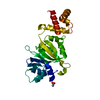
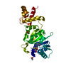
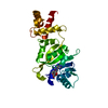
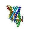
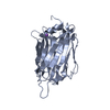
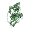
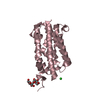
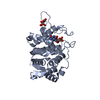
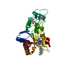
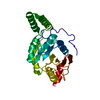
- PDB-1o9g: rRNA methyltransferase aviRa from Streptomyces viridochromogenes ... -
+ Open data

- Basic information
| Entry | Database: PDB / ID: 1o9g | ||||||
|---|---|---|---|---|---|---|---|
| Title | rRNA methyltransferase aviRa from Streptomyces viridochromogenes at 1.5A | ||||||
 Components Components | RRNA METHYLTRANSFERASE | ||||||
 Keywords Keywords |  TRANSFERASE / ANTIBIOTIC RESISTANCE / RRNA-METHYLTRANSFERASE / SE-MAD TRANSFERASE / ANTIBIOTIC RESISTANCE / RRNA-METHYLTRANSFERASE / SE-MAD | ||||||
| Function / homology |  Function and homology information23S rRNA (guanine2535-N1)-methyltransferase / rRNA (guanine-N1-)-methyltransferase activity / rRNA methylation / methylation / response to antibiotic Function and homology information23S rRNA (guanine2535-N1)-methyltransferase / rRNA (guanine-N1-)-methyltransferase activity / rRNA methylation / methylation / response to antibioticSimilarity search - Function | ||||||
| Biological species |  STREPTOMYCES VIRIDOCHROMOGENES (bacteria) STREPTOMYCES VIRIDOCHROMOGENES (bacteria) | ||||||
| Method | X-RAY DIFFRACTION / SYNCHROTRON / MAD / Resolution: 1.5 Å | ||||||
 Authors Authors | Mosbacher, T.G. / Schulz, G.E. | ||||||
 Citation Citation | Journal: J.Mol.Biol. / Year: 2003 Title: Crystal Structure of the Avilamycin Resistance-Conferring Methyltransferase Avira from Streptomyces Viridochromogenes Authors: Mosbacher, T.G. / Bechthold, A. / Schulz, G.E. | ||||||
| History |
|
- Structure visualization
| Structure viewer | Molecule: MolmilJmol/JSmol |
|---|
- Downloads & links
-Download
| PDBx/mmCIF format | 1o9g.cif.gz | 113.9 KB | Display | PDBx/mmCIF format |
|---|---|---|---|---|
| PDB format | pdb1o9g.ent.gz | 92.5 KB | Display | PDB format |
| PDBx/mmJSON format | 1o9g.json.gz | Tree view | PDBx/mmJSON format | |
| Others |  Other downloads Other downloads |
-Validation report
| Arichive directory | https://data.pdbj.org/pub/pdb/validation_reports/o9/1o9gftp://data.pdbj.org/pub/pdb/validation_reports/o9/1o9g | HTTPS FTP |
|---|
-Related structure data


-Links
 PDBj
PDBj- Assembly
| Deposited unit | 
| ||||||||
|---|---|---|---|---|---|---|---|---|---|
| 1 |
| ||||||||
| Unit cell |
|
-Components
| #1: Protein | Mass: 26833.492 Da / Num. of mol.: 1 / Mutation: YES Source method: isolated from a genetically manipulated source Source: (gene. exp.) STREPTOMYCES VIRIDOCHROMOGENES (bacteria)Plasmid: PRSETB / Production host: ESCHERICHIA COLI (E. coli) / Strain (production host): B834 / Variant (production host): SE-MET / References: UniProt: Q9F5K5 |
|---|---|
| #2: Water | ChemComp-HOH / Water Mass: 18.015 Da / Num. of mol.: 334 / Source method: isolated from a natural source / Formula: H2O Mass: 18.015 Da / Num. of mol.: 334 / Source method: isolated from a natural source / Formula: H2O |
| Compound details | ENGINEERED MUTATION ILE 11 MET, ARG 190 GLY AND LEU 239 MET. ALSO SEE REMARK 999 FOR MORE DETAILS ...ENGINEERED |
| Sequence details | SEQUENCE IN DATABASE INCORRECT FROM RESIDUE 180 TO 195. THE SWISS-PROT ACCESSION Q9F5K5 HAS THE ...SEQUENCE IN DATABASE INCORRECT FROM RESIDUE 180 TO 195. THE SWISS-PROT ACCESSION Q9F5K5 HAS THE SEQUENCE SARTGKGRCP |
-Experimental details
-Experiment
| Experiment | Method: X-RAY DIFFRACTION / Number of used crystals: 1 |
|---|
- Sample preparation
| Crystal | Density Matthews: 2 Å3/Da / Density % sol: 39 % | ||||||||||||||||||||||||
|---|---|---|---|---|---|---|---|---|---|---|---|---|---|---|---|---|---|---|---|---|---|---|---|---|---|
| Crystal grow | pH: 6.9 / Details: PEG 20000 9%, MES 6.6, pH 6.90 | ||||||||||||||||||||||||
| Crystal grow | *PLUS Temperature: 20 ℃ / Method: vapor diffusion, hanging drop / PH range low: 6.9 / PH range high: 6.6 | ||||||||||||||||||||||||
| Components of the solutions | *PLUS
|
-Data collection
| Diffraction | Mean temperature: 100 K |
|---|---|
| Diffraction source | Source: SYNCHROTRON / Site: EMBL/DESY, HAMBURG  / Beamline: BW7A / Wavelength: 0.9774 / Beamline: BW7A / Wavelength: 0.9774 |
| Detector | Date: Sep 8, 2002 |
| Radiation | Protocol: MAD / Monochromatic (M) / Laue (L): M / Scattering type: x-ray |
| Radiation wavelength | Wavelength: 0.9774 Å / Relative weight: 1 |
| Reflection | Resolution: 1.5→37.8 Å / Num. obs: 64711 / % possible obs: 97 % / Observed criterion σ(I): 2 / Redundancy: 4.3 % / Rsym value: 0.034 / Net I/σ(I): 38.7 |
| Reflection shell | Resolution: 1.5→1.52 Å / Redundancy: 3.4 % / Mean I/σ(I) obs: 6.6 / Rsym value: 0.2 / % possible all: 90 |
| Reflection | *PLUS Highest resolution: 1.5 Å / Lowest resolution: 15 Å / % possible obs: 97 % / Redundancy: 4.3 % / Num. measured all: 289006 / Rmerge(I) obs: 0.039 |
| Reflection shell | *PLUS % possible obs: 90 % / Redundancy: 3.4 % / Num. unique obs: 1482 / Rmerge(I) obs: 0.2 / Mean I/σ(I) obs: 6.6 |
- Processing
| Software |
| ||||||||||||||||||||
|---|---|---|---|---|---|---|---|---|---|---|---|---|---|---|---|---|---|---|---|---|---|
| Refinement | Method to determine structure: MAD / Resolution: 1.5→37.8 Å / SU B: 1.232 / SU ML: 0.047 / Cross valid method: THROUGHOUT / ESU R Free: 0.082 / Details: HYDROGENS HAVE BEEN ADDED IN THE RIDING POSITIONS
| ||||||||||||||||||||
| Displacement parameters | Biso mean: 9.096 Å2
| ||||||||||||||||||||
| Refinement step | Cycle: LAST / Resolution: 1.5→37.8 Å
| ||||||||||||||||||||
| Refinement | *PLUS Highest resolution: 1.5 Å / Lowest resolution: 25 Å / Num. reflection obs: 33212 | ||||||||||||||||||||
| Solvent computation | *PLUS | ||||||||||||||||||||
| Displacement parameters | *PLUS | ||||||||||||||||||||
| Refine LS restraints | *PLUS
|
