 Movie
Movie Controller


+ Open data

- Basic information
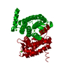
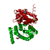
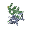
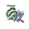
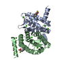
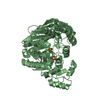
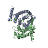
| Entry | Database: PDB / ID: 1nx0 | ||||||
|---|---|---|---|---|---|---|---|
| Title | Structure of Calpain Domain 6 in Complex with Calpastatin DIC | ||||||
 Components Components |
| ||||||
 Keywords Keywords | HYDROLASE/HYDROLASE INHIBITOR /  CALCIUM BINDING / HYDROLASE-HYDROLASE INHIBITOR complex CALCIUM BINDING / HYDROLASE-HYDROLASE INHIBITOR complex | ||||||
| Function / homology |  Function and homology information Function and homology informationcalcium-dependent cysteine-type endopeptidase inhibitor activity / Degradation of the extracellular matrix / calpain complex / calcium-dependent cysteine-type endopeptidase activity / cysteine-type endopeptidase inhibitor activity / protein catabolic process / calcium ion binding / proteolysis / plasma membrane / cytoplasmSimilarity search - Function | ||||||
| Biological species |  Sus scrofa (pig) Sus scrofa (pig) | ||||||
| Method | X-RAY DIFFRACTION / MOLECULAR REPLACEMENT / Resolution: 2.3 Å | ||||||
 Authors Authors | Todd, B. / Moore, D. / Deivanayagam, C.C.S. / Lin, G.-D. / Chattopadhyay, D. / Maki, M. / Wang, K.K.W. / Narayana, S.V.L. | ||||||
 Citation Citation | Journal: J.Mol.Biol. / Year: 2003 Title: A structural model for the inhibition of calpain by calpastatin: crystal structures of the native domain VI of calpain and its complexes with calpastatin peptide and a small molecule inhibitor. Authors: Todd, B. / Moore, D. / Deivanayagam, C.C.S. / Lin, G.-D. / Chattopadhyay, D. / Maki, M. / Wang, K.K.W. / Narayana, S.V.L. | ||||||
| History |
|
- Structure visualization
| Structure viewer | Molecule: MolmilJmol/JSmol |
|---|
- Downloads & links
-Download
| PDBx/mmCIF format | 1nx0.cif.gz | 91.4 KB | Display | PDBx/mmCIF format |
|---|---|---|---|---|
| PDB format | pdb1nx0.ent.gz | 68.5 KB | Display | PDB format |
| PDBx/mmJSON format | 1nx0.json.gz | Tree view | PDBx/mmJSON format | |
| Others |  Other downloads Other downloads |
-Validation report
| Arichive directory | https://data.pdbj.org/pub/pdb/validation_reports/nx/1nx0ftp://data.pdbj.org/pub/pdb/validation_reports/nx/1nx0 | HTTPS FTP |
|---|
-Related structure data


-Links
 PDBj
PDBj- Assembly
| Deposited unit | 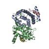
| ||||||||
|---|---|---|---|---|---|---|---|---|---|
| 1 |
| ||||||||
| Unit cell |
|
-Components
| #1: Protein | Mass: 19883.477 Da / Num. of mol.: 2 / Fragment: Domain VI Source method: isolated from a genetically manipulated source Source: (gene. exp.) Sus scrofa (pig) / Gene: CAPNS1 OR CAPN4 / Production host:  Escherichia coli (E. coli) / References: UniProt: P04574 Escherichia coli (E. coli) / References: UniProt: P04574#2: Protein/peptide | / calpain inhibitorMass: 1241.261 Da / Num. of mol.: 2 / Fragment: DIC, residues 230-241 Source method: isolated from a genetically manipulated source Source: (gene. exp.) Sus scrofa (pig) / Production host: Escherichia coli (E. coli) / References: UniProt: P49342, UniProt: P12675*PLUS#3: Protein/peptide | | Mass: 473.586 Da / Num. of mol.: 1 / Source method: obtained synthetically / Details: THE Peptide WAS CHEMICALLY SYNTHESIZED. #4: Chemical | ChemComp-CA /   Mass: 40.078 Da / Num. of mol.: 6 / Source method: obtained synthetically / Formula: Ca Mass: 40.078 Da / Num. of mol.: 6 / Source method: obtained synthetically / Formula: Ca#5: Water | ChemComp-HOH / | Water Mass: 18.015 Da / Num. of mol.: 150 / Source method: isolated from a natural source / Formula: H2O Mass: 18.015 Da / Num. of mol.: 150 / Source method: isolated from a natural source / Formula: H2O |
|---|
-Experimental details
-Experiment
| Experiment | Method: X-RAY DIFFRACTION / Number of used crystals: 1 |
|---|
- Sample preparation
| Crystal | Density Matthews: 2.42 Å3/Da / Density % sol: 49.08 % | ||||||||||||||||||||||||||||||||||||||||||||||||
|---|---|---|---|---|---|---|---|---|---|---|---|---|---|---|---|---|---|---|---|---|---|---|---|---|---|---|---|---|---|---|---|---|---|---|---|---|---|---|---|---|---|---|---|---|---|---|---|---|---|
| Crystal grow | Method: vapor diffusion, hanging drop / pH: 5.5 Details: PEG 6000, BME, EDTA, CaCl2, NaCl, pH 5.5, VAPOR DIFFUSION, HANGING DROP | ||||||||||||||||||||||||||||||||||||||||||||||||
| Crystal grow | *PLUS Method: vapor diffusion, hanging drop | ||||||||||||||||||||||||||||||||||||||||||||||||
| Components of the solutions | *PLUS
|
-Data collection
| Diffraction | Mean temperature: 103 K |
|---|---|
| Diffraction source | Source: ROTATING ANODE / Type: RIGAKU RUH3R / Wavelength: 1.5418 Å |
| Detector | Type: RIGAKU RAXIS IV / Detector: IMAGE PLATE |
| Radiation | Protocol: SINGLE WAVELENGTH / Monochromatic (M) / Laue (L): M / Scattering type: x-ray |
| Radiation wavelength | Wavelength: 1.5418 Å / Relative weight: 1 |
| Reflection | Resolution: 2.3→100 Å / Num. all: 18854 / Num. obs: 18854 / % possible obs: 98.2 % / Observed criterion σ(I): 0 / Rmerge(I) obs: 0.06 / Net I/σ(I): 17.5 |
| Reflection | *PLUS Highest resolution: 2.3 Å / Rmerge(I) obs: 0.06 |
- Processing
| Software |
| |||||||||||||||||||||||||
|---|---|---|---|---|---|---|---|---|---|---|---|---|---|---|---|---|---|---|---|---|---|---|---|---|---|---|
| Refinement | Method to determine structure: MOLECULAR REPLACEMENT / Resolution: 2.3→100 Å / σ(F): 3 / Stereochemistry target values: Engh & Huber
| |||||||||||||||||||||||||
| Refinement step | Cycle: LAST / Resolution: 2.3→100 Å
| |||||||||||||||||||||||||
| Refinement | *PLUS Highest resolution: 2.3 Å / Rfactor Rfree: 0.213 / Rfactor Rwork: 0.264 | |||||||||||||||||||||||||
| Solvent computation | *PLUS | |||||||||||||||||||||||||
| Displacement parameters | *PLUS | |||||||||||||||||||||||||
| Refine LS restraints | *PLUS
|
