 Movie
Movie Controller

[English] 日本語
 Yorodumi
- PDB-1nw2: The crystal structure of the mutant R82E of Thioredoxin from Alic... -
+ Open data

- Basic information
| Entry | Database: PDB / ID: 1nw2 | ||||||
|---|---|---|---|---|---|---|---|
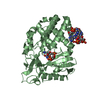
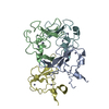
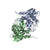
| Title | The crystal structure of the mutant R82E of Thioredoxin from Alicyclobacillus acidocaldarius | ||||||
 Components Components | THIOREDOXIN | ||||||
 Keywords Keywords | ELECTRON TRANSPORT / Thermostability / thioredoxin | ||||||
| Function / homology |  Function and homology information Function and homology information | ||||||
| Biological species |  Alicyclobacillus acidocaldarius (bacteria) Alicyclobacillus acidocaldarius (bacteria) | ||||||
| Method | X-RAY DIFFRACTION / SYNCHROTRON / MOLECULAR REPLACEMENT / Resolution: 1.9 Å | ||||||
 Authors Authors | Bartolucci, S. / De Simone, G. / Galdiero, S. / Improta, R. / Menchise, V. / Pedone, C. / Pedone, E. / Saviano, M. | ||||||
 Citation Citation | Journal: J.Bacteriol. / Year: 2003 Title: An integrated structural and computational study of the thermostability of two thioredoxin mutants from Alicyclobacillus acidocaldarius Authors: Bartolucci, S. / De Simone, G. / Galdiero, S. / Improta, R. / Menchise, V. / Pedone, C. / Pedone, E. / Saviano, M. #1: Journal: Biochem.J. / Year: 1999Title: Prediction and Experimental Testing of Bacillus acidocaldarius Thioredoxin Stability. Authors: Pedone, E. / Cannio, R. / Saviano, M. / Rossi, M. / Bartolucci, S. | ||||||
| History |
|
- Structure visualization
| Structure viewer | Molecule: MolmilJmol/JSmol |
|---|
- Downloads & links
-Download
| PDBx/mmCIF format | 1nw2.cif.gz | 187.8 KB | Display | PDBx/mmCIF format |
|---|---|---|---|---|
| PDB format | pdb1nw2.ent.gz | 149.9 KB | Display | PDB format |
| PDBx/mmJSON format | 1nw2.json.gz | Tree view | PDBx/mmJSON format | |
| Others |  Other downloads Other downloads |
-Validation report
| Arichive directory | https://data.pdbj.org/pub/pdb/validation_reports/nw/1nw2ftp://data.pdbj.org/pub/pdb/validation_reports/nw/1nw2 | HTTPS FTP |
|---|
-Related structure data
| Related structure data |  1nswC 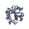 2trxS S: Starting model for refinement C: citing same article ( |
|---|---|
| Similar structure data |


-Links
 PDBj
PDBj


- Assembly
| Deposited unit | 
| ||||||||||
|---|---|---|---|---|---|---|---|---|---|---|---|
| 1 | 
| ||||||||||
| 2 | 
| ||||||||||
| 3 |
| ||||||||||
| 4 | 
| ||||||||||
| 5 | 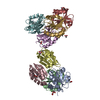
| ||||||||||
| 6 | 
| ||||||||||
| Unit cell |
|
-Components
| #1: Protein | / E.C.1.8.1.9 / TRX Mass: 11557.211 Da / Num. of mol.: 8 / Mutation: R82E Source method: isolated from a genetically manipulated source Source: (gene. exp.) Alicyclobacillus acidocaldarius (bacteria)Production host: Escherichia coli (E. coli)References: UniProt: P80579, thioredoxin-disulfide reductase#2: Chemical | ChemComp-ZN /   Mass: 65.409 Da / Num. of mol.: 9 / Source method: obtained synthetically / Formula: Zn Mass: 65.409 Da / Num. of mol.: 9 / Source method: obtained synthetically / Formula: Zn#3: Chemical | ChemComp-ACT / Acetate  Mass: 59.044 Da / Num. of mol.: 9 / Source method: obtained synthetically / Formula: C2H3O2 Mass: 59.044 Da / Num. of mol.: 9 / Source method: obtained synthetically / Formula: C2H3O2#4: Chemical | Cacodylic acid  Mass: 136.989 Da / Num. of mol.: 2 / Source method: obtained synthetically / Formula: C2H6AsO2 Mass: 136.989 Da / Num. of mol.: 2 / Source method: obtained synthetically / Formula: C2H6AsO2#5: Water | ChemComp-HOH / | Water Mass: 18.015 Da / Num. of mol.: 667 / Source method: isolated from a natural source / Formula: H2O Mass: 18.015 Da / Num. of mol.: 667 / Source method: isolated from a natural source / Formula: H2O |
|---|
-Experimental details
-Experiment
| Experiment | Method: X-RAY DIFFRACTION / Number of used crystals: 1 |
|---|
- Sample preparation
| Crystal | Density Matthews: 1.97 Å3/Da / Density % sol: 37 % | ||||||||||||||||||||||||||||||
|---|---|---|---|---|---|---|---|---|---|---|---|---|---|---|---|---|---|---|---|---|---|---|---|---|---|---|---|---|---|---|---|
| Crystal grow | Temperature: 295 K / Method: vapor diffusion, hanging drop / pH: 8 Details: Peg 8000, calcium acetate, CACODYLATE, VAPOR DIFFUSION, HANGING DROP, temperature 295K | ||||||||||||||||||||||||||||||
| Crystal grow | *PLUS Temperature: 22 ℃ / Method: vapor diffusion, hanging drop | ||||||||||||||||||||||||||||||
| Components of the solutions | *PLUS
|
-Data collection
| Diffraction | Mean temperature: 100 K |
|---|---|
| Diffraction source | Source: SYNCHROTRON / Site: EMBL/DESY, HAMBURG  / Beamline: X11 / Wavelength: 0.9072 Å / Beamline: X11 / Wavelength: 0.9072 Å |
| Detector | Type: MARRESEARCH / Detector: IMAGE PLATE |
| Radiation | Protocol: SINGLE WAVELENGTH / Monochromatic (M) / Laue (L): M / Scattering type: x-ray |
| Radiation wavelength | Wavelength: 0.9072 Å / Relative weight: 1 |
| Reflection | Resolution: 1.9→20 Å / Num. all: 52158 / Num. obs: 52158 / % possible obs: 84.1 % / Observed criterion σ(I): 0 / Rsym value: 0.051 / Net I/σ(I): 20.1 |
| Reflection shell | Resolution: 1.9→1.97 Å / Mean I/σ(I) obs: 4.3 / Rsym value: 0.262 / % possible all: 85.7 |
| Reflection | *PLUS Num. measured all: 234705 / Rmerge(I) obs: 0.051 |
| Reflection shell | *PLUS % possible obs: 85.7 % / Rmerge(I) obs: 0.262 |
- Processing
| Software |
| |||||||||||||||||||||||||
|---|---|---|---|---|---|---|---|---|---|---|---|---|---|---|---|---|---|---|---|---|---|---|---|---|---|---|
| Refinement | Method to determine structure: MOLECULAR REPLACEMENT Starting model: PDB ENTRY 2TRX Resolution: 1.9→20 Å / Cross valid method: THROUGHOUT / σ(F): 0 / Stereochemistry target values: Engh & Huber
| |||||||||||||||||||||||||
| Refinement step | Cycle: LAST / Resolution: 1.9→20 Å
| |||||||||||||||||||||||||
| Refine LS restraints |
| |||||||||||||||||||||||||
| Refinement | *PLUS % reflection Rfree: 5 % | |||||||||||||||||||||||||
| Solvent computation | *PLUS | |||||||||||||||||||||||||
| Displacement parameters | *PLUS |
