 Movie
Movie Controller

[English] 日本語
 Yorodumi
- PDB-2pmv: Crystal Structure of Human Intrinsic Factor- Cobalamin Complex at... -
+ Open data

- Basic information
| Entry | Database: PDB / ID: 2pmv | |||||||||
|---|---|---|---|---|---|---|---|---|---|---|
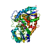
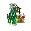
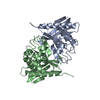
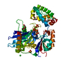
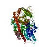
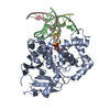
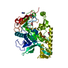
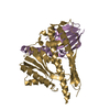
| Title | Crystal Structure of Human Intrinsic Factor- Cobalamin Complex at 2.6 A Resolution | |||||||||
 Components Components | Gastric intrinsic factor | |||||||||
 Keywords Keywords |  TRANSPORT PROTEIN / Cobalamin transport protein Alpha6-Alpha6 motif two domain protein TRANSPORT PROTEIN / Cobalamin transport protein Alpha6-Alpha6 motif two domain protein | |||||||||
| Function / homology |  Function and homology information Function and homology informationDefective CBLIF causes IFD / Defective AMN causes MGA1 / Defective CUBN causes MGA1 / cargo receptor ligand activity / Uptake of dietary cobalamins into enterocytes / cobalt ion transport / cobalamin transport / cobalamin binding / microvillus / lysosomal lumen ...Defective CBLIF causes IFD / Defective AMN causes MGA1 / Defective CUBN causes MGA1 / cargo receptor ligand activity / Uptake of dietary cobalamins into enterocytes / cobalt ion transport / cobalamin transport / cobalamin binding / microvillus / lysosomal lumen / endosome / apical plasma membrane / extracellular space / extracellular regionSimilarity search - Function | |||||||||
| Biological species |  Homo sapiens (human) Homo sapiens (human) | |||||||||
| Method | X-RAY DIFFRACTION / SYNCHROTRON / SAD / Resolution: 2.6 Å | |||||||||
 Authors Authors | Mathews, F.S. / Gordon, M.M. / Chen, Z. / Rajashankar, K.R. / Ealick, S.E. / Alpers, D.H. / Sukumar, N. | |||||||||
 Citation Citation | Journal: Proc.Natl.Acad.Sci.USA / Year: 2007 Title: Crystal structure of human intrinsic factor: Cobalamin complex at 2.6-A resolution Authors: Mathews, F.S. / Gordon, M.M. / Chen, Z. / Rajashankar, K.R. / Ealick, S.E. / Alpers, D.H. / Sukumar, N. | |||||||||
| History |
|
- Structure visualization
| Structure viewer | Molecule: MolmilJmol/JSmol |
|---|
- Downloads & links
-Download
| PDBx/mmCIF format | 2pmv.cif.gz | 263.8 KB | Display | PDBx/mmCIF format |
|---|---|---|---|---|
| PDB format | pdb2pmv.ent.gz | 216.4 KB | Display | PDB format |
| PDBx/mmJSON format | 2pmv.json.gz | Tree view | PDBx/mmJSON format | |
| Others |  Other downloads Other downloads |
-Validation report
| Arichive directory | https://data.pdbj.org/pub/pdb/validation_reports/pm/2pmvftp://data.pdbj.org/pub/pdb/validation_reports/pm/2pmv | HTTPS FTP |
|---|
-Related structure data
| Similar structure data |
|---|


-Links
 PDBj
PDBj- Assembly
| Deposited unit | 
| ||||||||
|---|---|---|---|---|---|---|---|---|---|
| 1 | 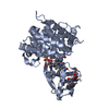
| ||||||||
| 2 | 
| ||||||||
| 3 | 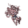
| ||||||||
| 4 | 
| ||||||||
| Unit cell |
| ||||||||
| Details | The biological assembly is a monomer. |
-Components
| #1: Protein | Mass: 43448.379 Da / Num. of mol.: 4 Source method: isolated from a genetically manipulated source Source: (gene. exp.) Homo sapiens (human) / Production host:  Pichia pastoris (fungus) / References: UniProt: P27352 Pichia pastoris (fungus) / References: UniProt: P27352#2: Polysaccharide | / Mass: 424.401 Da / Num. of mol.: 2Source method: isolated from a genetically manipulated source #3: Chemical | Vitamin B12  Mass: 1330.356 Da / Num. of mol.: 2 / Source method: obtained synthetically / Formula: C62H89CoN13O14P Mass: 1330.356 Da / Num. of mol.: 2 / Source method: obtained synthetically / Formula: C62H89CoN13O14P#4: Water | ChemComp-HOH / | Water Mass: 18.015 Da / Num. of mol.: 459 / Source method: isolated from a natural source / Formula: H2O Mass: 18.015 Da / Num. of mol.: 459 / Source method: isolated from a natural source / Formula: H2O |
|---|
-Experimental details
-Experiment
| Experiment | Method: X-RAY DIFFRACTION / Number of used crystals: 1 |
|---|
- Sample preparation
| Crystal | Density Matthews: 2.56 Å3/Da / Density % sol: 51.9 % |
|---|---|
| Crystal grow | Temperature: 295 K / Method: vapor diffusion, sitting drop / pH: 6 Details: 10% PEG20000, 100mM MES, 20mM CaCL2 and 9mM BaCL2, pH 6.0, VAPOR DIFFUSION, SITTING DROP, temperature 295K |
-Data collection
| Diffraction | Mean temperature: 100 K |
|---|---|
| Diffraction source | Source: SYNCHROTRON / Site: APS  / Beamline: 8-BM / Wavelength: 1.6059 Å / Beamline: 8-BM / Wavelength: 1.6059 Å |
| Detector | Type: ADSC QUANTUM 315 / Detector: CCD |
| Radiation | Protocol: SINGLE WAVELENGTH / Monochromatic (M) / Laue (L): M / Scattering type: x-ray |
| Radiation wavelength | Wavelength: 1.6059 Å / Relative weight: 1 |
| Reflection | Resolution: 2.5→50 Å / Num. all: 61149 / Num. obs: 55707 / % possible obs: 91.1 % / Observed criterion σ(F): -3 / Observed criterion σ(I): -3 / Redundancy: 3.5 % / Biso Wilson estimate: 36.5 Å2 / Rmerge(I) obs: 0.07 / Net I/σ(I): 16.5 |
| Reflection shell | Resolution: 2.5→2.6 Å / Redundancy: 3.2 % / Rmerge(I) obs: 0.491 / Mean I/σ(I) obs: 2.2 / % possible all: 54.7 |
- Processing
| Software |
| |||||||||||||||||||||||||
|---|---|---|---|---|---|---|---|---|---|---|---|---|---|---|---|---|---|---|---|---|---|---|---|---|---|---|
| Refinement | Method to determine structure: SAD / Resolution: 2.6→37.53 Å / Rfactor Rfree error: 0.006 / Data cutoff high absF: 491016.14 / Data cutoff low absF: 0 / Isotropic thermal model: RESTRAINED / Cross valid method: THROUGHOUT / σ(F): 0 / Stereochemistry target values: Engh & Huber
| |||||||||||||||||||||||||
| Displacement parameters | Biso mean: 56 Å2 | |||||||||||||||||||||||||
| Refine analyze |
| |||||||||||||||||||||||||
| Refinement step | Cycle: LAST / Resolution: 2.6→37.53 Å
| |||||||||||||||||||||||||
| Refine LS restraints |
| |||||||||||||||||||||||||
| LS refinement shell | Resolution: 2.6→2.76 Å / Rfactor Rfree error: 0.023 / Total num. of bins used: 6
|
