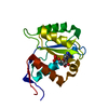
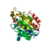
 Movie
Movie Controller

[English] 日本語
 Yorodumi
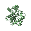
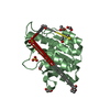
- PDB-1nqz: The structure of a CoA pyrophosphatase from D. Radiodurans comple... -
+ Open data

- Basic information
| Entry | Database: PDB / ID: 1nqz | ||||||
|---|---|---|---|---|---|---|---|
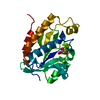
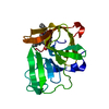
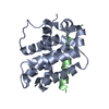
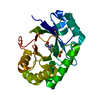
| Title | The structure of a CoA pyrophosphatase from D. Radiodurans complexed with a magnesium ion | ||||||
 Components Components | CoA pyrophosphatase (MutT/nudix family protein) | ||||||
 Keywords Keywords |  HYDROLASE / Nudix / MutT / pyrophosphatase / CoA / D.Radiodurans HYDROLASE / Nudix / MutT / pyrophosphatase / CoA / D.Radiodurans | ||||||
| Function / homology |  Function and homology information Function and homology informationcoenzyme A diphosphatase activity / coenzyme A catabolic process / Hydrolases; Acting on acid anhydrides; In phosphorus-containing anhydrides / magnesium ion bindingSimilarity search - Function | ||||||
| Biological species |  Deinococcus radiodurans (radioresistant) Deinococcus radiodurans (radioresistant) | ||||||
| Method | X-RAY DIFFRACTION / SYNCHROTRON / MOLECULAR REPLACEMENT / Resolution: 1.7 Å | ||||||
 Authors Authors | Kang, L.W. / Gabelli, S.B. / Bianchet, M.A. / Xu, W.L. / Bessman, M.J. / Amzel, L.M. | ||||||
 Citation Citation | Journal: J.Bacteriol. / Year: 2003 Title: Structure of a coenzyme A pyrophosphatase from Deinococcus radiodurans: a member of the Nudix family. Authors: Kang, L.W. / Gabelli, S.B. / Bianchet, M.A. / Xu, W.L. / Bessman, M.J. / Amzel, L.M. | ||||||
| History |
|
- Structure visualization
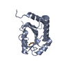
| Structure viewer | Molecule: MolmilJmol/JSmol |
|---|
- Downloads & links
-Download
| PDBx/mmCIF format | 1nqz.cif.gz | 48.7 KB | Display | PDBx/mmCIF format |
|---|---|---|---|---|
| PDB format | pdb1nqz.ent.gz | 33.4 KB | Display | PDB format |
| PDBx/mmJSON format | 1nqz.json.gz | Tree view | PDBx/mmJSON format | |
| Others |  Other downloads Other downloads |
-Validation report
| Arichive directory | https://data.pdbj.org/pub/pdb/validation_reports/nq/1nqzftp://data.pdbj.org/pub/pdb/validation_reports/nq/1nqz | HTTPS FTP |
|---|
-Related structure data
| Related structure data | 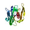 1nqySC S: Starting model for refinement C: citing same article ( |
|---|---|
| Similar structure data |

-Links
 PDBj
PDBj

- Assembly
| Deposited unit | 
| ||||||||
|---|---|---|---|---|---|---|---|---|---|
| 1 |
| ||||||||
| Unit cell |
| ||||||||
| Components on special symmetry positions |
|
-Components
| #1: Protein | Mass: 21273.229 Da / Num. of mol.: 1 Source method: isolated from a genetically manipulated source Source: (gene. exp.) Deinococcus radiodurans (radioresistant)Gene: dr1184 / Production host: Escherichia coli (E. coli) / References: UniProt: Q9RV46, nucleotide diphosphatase |
|---|---|
| #2: Chemical | ChemComp-MG /   Mass: 24.305 Da / Num. of mol.: 1 / Source method: obtained synthetically / Formula: Mg Mass: 24.305 Da / Num. of mol.: 1 / Source method: obtained synthetically / Formula: Mg |
| #3: Water | ChemComp-HOH / Water Mass: 18.015 Da / Num. of mol.: 123 / Source method: isolated from a natural source / Formula: H2O Mass: 18.015 Da / Num. of mol.: 123 / Source method: isolated from a natural source / Formula: H2O |
-Experimental details
-Experiment
| Experiment | Method: X-RAY DIFFRACTION / Number of used crystals: 1 |
|---|
- Sample preparation
| Crystal | Density Matthews: 1.96 Å3/Da / Density % sol: 37.21 % | ||||||||||||||||||||||||||||||||||||
|---|---|---|---|---|---|---|---|---|---|---|---|---|---|---|---|---|---|---|---|---|---|---|---|---|---|---|---|---|---|---|---|---|---|---|---|---|---|
| Crystal grow | Temperature: 293 K / Method: vapor diffusion, hanging drop / pH: 6 Details: PEG1500, MES buffer, pH 6.0, VAPOR DIFFUSION, HANGING DROP, temperature 293K | ||||||||||||||||||||||||||||||||||||
| Crystal grow | *PLUS Temperature: 20 ℃ / pH: 7.5 / Method: vapor diffusion, sitting drop | ||||||||||||||||||||||||||||||||||||
| Components of the solutions | *PLUS
|
-Data collection
| Diffraction | Mean temperature: 90 K |
|---|---|
| Diffraction source | Source: SYNCHROTRON / Site: NSLS  / Beamline: X25 / Wavelength: 1.009 Å / Beamline: X25 / Wavelength: 1.009 Å |
| Detector | Type: ADSC QUANTUM 4 / Detector: CCD / Date: Oct 20, 2002 |
| Radiation | Protocol: SINGLE WAVELENGTH / Monochromatic (M) / Laue (L): M / Scattering type: x-ray |
| Radiation wavelength | Wavelength: 1.009 Å / Relative weight: 1 |
| Reflection | Resolution: 1.7→30 Å / Num. obs: 19112 / % possible obs: 99.6 % / Observed criterion σ(F): 0 / Observed criterion σ(I): 0 / Rsym value: 0.062 / Net I/σ(I): 15.4 |
| Reflection | *PLUS Lowest resolution: 30 Å / Num. measured all: 100105 / Rmerge(I) obs: 0.062 |
| Reflection shell | *PLUS % possible obs: 97.5 % / Mean I/σ(I) obs: 5.5 |
- Processing
| Software |
| ||||||||||||||||||||
|---|---|---|---|---|---|---|---|---|---|---|---|---|---|---|---|---|---|---|---|---|---|
| Refinement | Method to determine structure: MOLECULAR REPLACEMENT Starting model: 1NQY Resolution: 1.7→30 Å / σ(F): 0 / Stereochemistry target values: Engh & Huber
| ||||||||||||||||||||
| Refinement step | Cycle: LAST / Resolution: 1.7→30 Å
| ||||||||||||||||||||
| Refine LS restraints |
| ||||||||||||||||||||
| Refinement | *PLUS Lowest resolution: 30 Å / % reflection Rfree: 10 % | ||||||||||||||||||||
| Solvent computation | *PLUS | ||||||||||||||||||||
| Displacement parameters | *PLUS | ||||||||||||||||||||
| LS refinement shell | *PLUS Rfactor Rfree: 0.325 / Rfactor Rwork: 0.313 |
