 Movie
Movie Controller

[English] 日本語
 Yorodumi
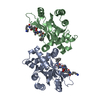
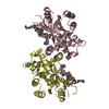
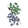
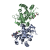
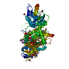
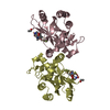
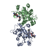
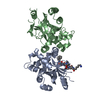
- PDB-1njt: COMPLEX STRUCTURE OF HCMV PROTEASE AND A PEPTIDOMIMETIC INHIBITOR -
+ Open data

- Basic information
| Entry | Database: PDB / ID: 1njt | ||||||
|---|---|---|---|---|---|---|---|
| Title | COMPLEX STRUCTURE OF HCMV PROTEASE AND A PEPTIDOMIMETIC INHIBITOR | ||||||
 Components Components |
| ||||||
 Keywords Keywords | HYDROLASE/HYDROLASE INHIBITOR /  PROTEASE / PEPTIDOMIMETIC INHIBITOR / INDUCED-FIT / HYDROLASE-HYDROLASE INHIBITOR COMPLEX PROTEASE / PEPTIDOMIMETIC INHIBITOR / INDUCED-FIT / HYDROLASE-HYDROLASE INHIBITOR COMPLEX | ||||||
| Function / homology |  Function and homology informationassemblin / viral release from host cell / host cell cytoplasm / serine-type endopeptidase activity / host cell nucleus / proteolysis / identical protein binding Function and homology informationassemblin / viral release from host cell / host cell cytoplasm / serine-type endopeptidase activity / host cell nucleus / proteolysis / identical protein bindingSimilarity search - Function | ||||||
| Biological species |   Human herpesvirus 5 Human herpesvirus 5 | ||||||
| Method | X-RAY DIFFRACTION / SYNCHROTRON / MOLECULAR REPLACEMENT / Resolution: 2.5 Å | ||||||
 Authors Authors | Khayat, R. / Batra, R. / Qian, C. / Halmos, T. / Bailey, M. / Tong, L. | ||||||
 Citation Citation | Journal: Biochemistry / Year: 2003 Title: Structural and Biochemical Studies of Inhibitor Binding to Human Cytomegalovirus Protease Authors: Khayat, R. / Batra, R. / Qian, C. / Halmos, T. / Bailey, M. / Tong, L. | ||||||
| History |
|
- Structure visualization
| Structure viewer | Molecule: MolmilJmol/JSmol |
|---|
- Downloads & links
-Download
| PDBx/mmCIF format | 1njt.cif.gz | 192.2 KB | Display | PDBx/mmCIF format |
|---|---|---|---|---|
| PDB format | pdb1njt.ent.gz | 157.8 KB | Display | PDB format |
| PDBx/mmJSON format | 1njt.json.gz | Tree view | PDBx/mmJSON format | |
| Others |  Other downloads Other downloads |
-Validation report
| Arichive directory | https://data.pdbj.org/pub/pdb/validation_reports/nj/1njtftp://data.pdbj.org/pub/pdb/validation_reports/nj/1njt | HTTPS FTP |
|---|
-Related structure data


-Links
 PDBj
PDBj- Assembly
| Deposited unit | 
| ||||||||
|---|---|---|---|---|---|---|---|---|---|
| 1 | 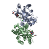
| ||||||||
| 2 | 
| ||||||||
| Unit cell |
|
-Components
| #1: Protein | / Protease / Capsid assembly protein Mass: 28130.547 Da / Num. of mol.: 4 / Fragment: Assemblin / Mutation: A143Q Source method: isolated from a genetically manipulated source Source: (gene. exp.) Human herpesvirus 5 / Genus: Cytomegalovirus / Gene: UL80 OR APNG / Production host:  Escherichia coli (E. coli) / References: UniProt: P16753, assemblin Escherichia coli (E. coli) / References: UniProt: P16753, assemblin#2: Protein/peptide |   Type: Peptide-like / Class: Inhibitor / Mass: 551.557 Da / Num. of mol.: 4 / Source method: obtained synthetically Type: Peptide-like / Class: Inhibitor / Mass: 551.557 Da / Num. of mol.: 4 / Source method: obtained syntheticallyDetails: Residue peptide of the peptidomimetic inhibitor was chemically synthesized. References: N-acetyl-L-valyl-3,3-dimethyl-L-alpha-aspartyl-N~4~,N~4~-dimethyl-N~1~-[(1R)-3,3,3-trifluoro-1-methyl-2-oxopropyl]-L-as partamide #3: Chemical | ChemComp-CL / Chloride  Mass: 35.453 Da / Num. of mol.: 4 / Source method: obtained synthetically / Formula: Cl Mass: 35.453 Da / Num. of mol.: 4 / Source method: obtained synthetically / Formula: Cl#4: Water | ChemComp-HOH / | Water Mass: 18.015 Da / Num. of mol.: 243 / Source method: isolated from a natural source / Formula: H2O Mass: 18.015 Da / Num. of mol.: 243 / Source method: isolated from a natural source / Formula: H2O |
|---|
-Experimental details
-Experiment
| Experiment | Method: X-RAY DIFFRACTION / Number of used crystals: 1 |
|---|
- Sample preparation
| Crystal | Density Matthews: 2.63 Å3/Da / Density % sol: 53.25 % | |||||||||||||||||||||||||||||||||||||||||||||||||||||||||||||||||||||||||||||||||||||||||||||||||||||||||
|---|---|---|---|---|---|---|---|---|---|---|---|---|---|---|---|---|---|---|---|---|---|---|---|---|---|---|---|---|---|---|---|---|---|---|---|---|---|---|---|---|---|---|---|---|---|---|---|---|---|---|---|---|---|---|---|---|---|---|---|---|---|---|---|---|---|---|---|---|---|---|---|---|---|---|---|---|---|---|---|---|---|---|---|---|---|---|---|---|---|---|---|---|---|---|---|---|---|---|---|---|---|---|---|---|---|---|
| Crystal grow | Temperature: 293 K / Method: vapor diffusion, hanging drop / pH: 7 Details: PEG 4000, HEPES, EDTA, Sodium Chloride, Sodium Sulfate, DTT, Spermine_HCl, Glycerol, pH 7.0, VAPOR DIFFUSION, HANGING DROP, temperature 293K | |||||||||||||||||||||||||||||||||||||||||||||||||||||||||||||||||||||||||||||||||||||||||||||||||||||||||
| Crystal grow | *PLUS Temperature: 21 ℃ | |||||||||||||||||||||||||||||||||||||||||||||||||||||||||||||||||||||||||||||||||||||||||||||||||||||||||
| Components of the solutions | *PLUS
|
-Data collection
| Diffraction | Mean temperature: 100 K |
|---|---|
| Diffraction source | Source: SYNCHROTRON / Site: NSLS  / Beamline: X12C / Wavelength: 0.9797 Å / Beamline: X12C / Wavelength: 0.9797 Å |
| Detector | Type: CUSTOM-MADE / Detector: CCD / Date: Aug 13, 2001 |
| Radiation | Monochromator: Si (111) / Protocol: SINGLE WAVELENGTH / Monochromatic (M) / Laue (L): M / Scattering type: x-ray |
| Radiation wavelength | Wavelength: 0.9797 Å / Relative weight: 1 |
| Reflection | Resolution: 2.5→50 Å / Num. all: 43637 / Num. obs: 43637 / % possible obs: 85 % / Observed criterion σ(I): 0 / Biso Wilson estimate: 20.2 Å2 |
| Reflection shell | Resolution: 2.5→2.63 Å / % possible all: 72.7 |
| Reflection | *PLUS Highest resolution: 2.5 Å / Lowest resolution: 50 Å / % possible obs: 85 % / Num. measured all: 182204 / Rmerge(I) obs: 0.06 |
- Processing
| Software |
| ||||||||||||||||||||||||||||||||||||
|---|---|---|---|---|---|---|---|---|---|---|---|---|---|---|---|---|---|---|---|---|---|---|---|---|---|---|---|---|---|---|---|---|---|---|---|---|---|
| Refinement | Method to determine structure: MOLECULAR REPLACEMENT / Resolution: 2.5→19.95 Å / Rfactor Rfree error: 0.005 / Data cutoff high absF: 441167.76 / Data cutoff high rms absF: 441167.76 / Data cutoff low absF: 0 / Isotropic thermal model: RESTRAINED / Cross valid method: THROUGHOUT / σ(F): 2 / Stereochemistry target values: Engh & Huber
| ||||||||||||||||||||||||||||||||||||
| Solvent computation | Solvent model: FLAT MODEL / Bsol: 23.9752 Å2 / ksol: 0.336613 e/Å3 | ||||||||||||||||||||||||||||||||||||
| Displacement parameters | Biso mean: 28.1 Å2
| ||||||||||||||||||||||||||||||||||||
| Refine analyze |
| ||||||||||||||||||||||||||||||||||||
| Refinement step | Cycle: LAST / Resolution: 2.5→19.95 Å
| ||||||||||||||||||||||||||||||||||||
| Refine LS restraints |
| ||||||||||||||||||||||||||||||||||||
| LS refinement shell | Resolution: 2.5→2.66 Å / Rfactor Rfree error: 0.02 / Total num. of bins used: 6
| ||||||||||||||||||||||||||||||||||||
| Xplor file |
| ||||||||||||||||||||||||||||||||||||
| Refinement | *PLUS Highest resolution: 2.5 Å / Lowest resolution: 20 Å / Rfactor Rfree: 0.273 | ||||||||||||||||||||||||||||||||||||
| Solvent computation | *PLUS | ||||||||||||||||||||||||||||||||||||
| Displacement parameters | *PLUS | ||||||||||||||||||||||||||||||||||||
| Refine LS restraints | *PLUS
|
