 Movie
Movie Controller


+ Open data

- Basic information
| Entry | Database: PDB / ID: 1n9j | ||||||
|---|---|---|---|---|---|---|---|
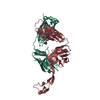
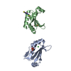
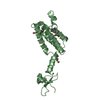
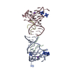
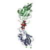
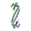
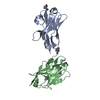
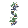
| Title | Solution Structure of the 3D domain swapped dimer of Stefin A | ||||||
 Components Components | Cystatin A | ||||||
 Keywords Keywords | HYDROLASE INHIBITOR / domain swapped / stefin A / cystatins / amyloid | ||||||
| Function / homology |  Function and homology information Function and homology informationnegative regulation of peptidase activity / peptidase inhibitor complex / Formation of the cornified envelope / peptide cross-linking / cornified envelope / cysteine-type endopeptidase inhibitor activity / keratinocyte differentiation / negative regulation of proteolysis / cell-cell adhesion / protease binding ...negative regulation of peptidase activity / peptidase inhibitor complex / Formation of the cornified envelope / peptide cross-linking / cornified envelope / cysteine-type endopeptidase inhibitor activity / keratinocyte differentiation / negative regulation of proteolysis / cell-cell adhesion / protease binding / extracellular space / nucleoplasm / cytosol / cytoplasmSimilarity search - Function | ||||||
| Biological species |  Homo sapiens (human) Homo sapiens (human) | ||||||
| Method | SOLUTION NMR / simulated annealing | ||||||
| Model type details | minimized average | ||||||
 Authors Authors | Staniforth, R.A. / Giannini, S. / Higgins, L.D. / Conroy, M.J. / Hounslow, A.M. / Jerala, R. / Craven, C.J. / Waltho, J.P. | ||||||
 Citation Citation | Journal: Embo J. / Year: 2001 Title: Three-dimensional domain swapping in the folded and molten-globule states of cystatins, an amyloid-forming structural superfamily Authors: Staniforth, R.A. / Giannini, S. / Higgins, L.D. / Conroy, M.J. / Hounslow, A.M. / Jerala, R. / Craven, C.J. / Waltho, J.P. | ||||||
| History |
|
- Structure visualization
| Structure viewer | Molecule: MolmilJmol/JSmol |
|---|
- Downloads & links
-Download
| PDBx/mmCIF format | 1n9j.cif.gz | 73 KB | Display | PDBx/mmCIF format |
|---|---|---|---|---|
| PDB format | pdb1n9j.ent.gz | 56.3 KB | Display | PDB format |
| PDBx/mmJSON format | 1n9j.json.gz | Tree view | PDBx/mmJSON format | |
| Others |  Other downloads Other downloads |
-Validation report
| Arichive directory | https://data.pdbj.org/pub/pdb/validation_reports/n9/1n9jftp://data.pdbj.org/pub/pdb/validation_reports/n9/1n9j | HTTPS FTP |
|---|
-Related structure data
| Related structure data | |
|---|---|
| Similar structure data |


-Links
 PDBj
PDBj

- Assembly
| Deposited unit | 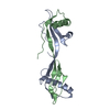
| |||||||||
|---|---|---|---|---|---|---|---|---|---|---|
| 1 |
| |||||||||
| NMR ensembles |
|
-Components
| #1: Protein | / Stefin A / Cystatin AS Mass: 11020.464 Da / Num. of mol.: 2 Source method: isolated from a genetically manipulated source Source: (gene. exp.) Homo sapiens (human) / Plasmid: pET3A / Species (production host): Escherichia coli / Production host:  Escherichia coli BL21(DE3) (bacteria) / Strain (production host): BL21-DE3 / References: UniProt: P01040 Escherichia coli BL21(DE3) (bacteria) / Strain (production host): BL21-DE3 / References: UniProt: P01040 |
|---|
-Experimental details
-Experiment
| Experiment | Method: SOLUTION NMR | ||||||||||||||||||||||||||||
|---|---|---|---|---|---|---|---|---|---|---|---|---|---|---|---|---|---|---|---|---|---|---|---|---|---|---|---|---|---|
| NMR experiment |
|
- Sample preparation
| Details |
| |||||||||
|---|---|---|---|---|---|---|---|---|---|---|
| Sample conditions | pH: 5.5 / Pressure: ambient / Temperature: 308 K | |||||||||
| Crystal grow | *PLUS Method: other / Details: NMR |
-NMR measurement
| Radiation | Protocol: SINGLE WAVELENGTH / Monochromatic (M) / Laue (L): M | |||||||||||||||
|---|---|---|---|---|---|---|---|---|---|---|---|---|---|---|---|---|
| Radiation wavelength | Relative weight: 1 | |||||||||||||||
| NMR spectrometer |
|
- Processing
| NMR software |
| ||||||||||||||||
|---|---|---|---|---|---|---|---|---|---|---|---|---|---|---|---|---|---|
| Refinement | Method: simulated annealing / Software ordinal: 1 Details: Distance restraints (NOEs and hydrogen bonds) were taken from the data used for the stefin A monomer structure (Martin et al., 1995), except those involving residues V48-G50 and N77-L80 ...Details: Distance restraints (NOEs and hydrogen bonds) were taken from the data used for the stefin A monomer structure (Martin et al., 1995), except those involving residues V48-G50 and N77-L80 where chemical shift perturbation was observed following resonance assignment. The NOEs were specified as intermolecular and intramolecular according to domain-swapped topology inferred from isotope filtering experiments. An additional hydrogen bond restraint was included between 48 and 50 based on evidence from protection experiments on the dimer (Jerala and Zerovnik, 1999). Dihedral restraints (phi and psi) were determined using the 1H-alpha, 15N, 13C-alpha, 13C-beta and 13C' chemical shifts and the program TALOS (Cornilescu et al., 1999). Where TALOS gave a `poor' match, the experimental phi dihedral angle was taken from the data used for the stefin A monomer structure calculation. | ||||||||||||||||
| NMR representative | Selection criteria: minimized average structure | ||||||||||||||||
| NMR ensemble | Conformers submitted total number: 1 |
