 Movie
Movie Controller

[English] 日本語
 Yorodumi
- PDB-1n8w: Biochemical and Structural Studies of Malate Synthase from Mycoba... -
+ Open data

- Basic information
| Entry | Database: PDB / ID: 1n8w | ||||||
|---|---|---|---|---|---|---|---|
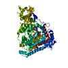
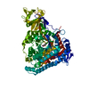
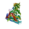
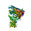
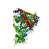
| Title | Biochemical and Structural Studies of Malate Synthase from Mycobacterium tuberculosis | ||||||
 Components Components | Probable malate synthase G | ||||||
 Keywords Keywords |  LYASE / MALATE SYNTHASE / GLYOXYLATE PATHWAY / MYCOBACTERIUM TUBERCULOSIS / MALATE / COENZYME A / GLCB / GLYOXYLATE / Structural Genomics / PSI / Protein Structure Initiative / TB Structural Genomics Consortium / TBSGC LYASE / MALATE SYNTHASE / GLYOXYLATE PATHWAY / MYCOBACTERIUM TUBERCULOSIS / MALATE / COENZYME A / GLCB / GLYOXYLATE / Structural Genomics / PSI / Protein Structure Initiative / TB Structural Genomics Consortium / TBSGC | ||||||
| Function / homology |  Function and homology information Function and homology informationhost cell extracellular matrix binding / capsule / malate synthase / malate synthase activity / coenzyme A binding / adhesion of symbiont to host / coenzyme A metabolic process / glyoxylate catabolic process / glyoxylate cycle / fibronectin binding ...host cell extracellular matrix binding / capsule / malate synthase / malate synthase activity / coenzyme A binding / adhesion of symbiont to host / coenzyme A metabolic process / glyoxylate catabolic process / glyoxylate cycle / fibronectin binding / laminin binding / tricarboxylic acid cycle / peptidoglycan-based cell wall / manganese ion binding / magnesium ion binding / cell surface / protein homodimerization activity / extracellular region / plasma membrane / cytosol / cytoplasmSimilarity search - Function | ||||||
| Biological species |   Mycobacterium tuberculosis (bacteria) Mycobacterium tuberculosis (bacteria) | ||||||
| Method | X-RAY DIFFRACTION / SYNCHROTRON / MOLECULAR REPLACEMENT / Resolution: 2.7 Å | ||||||
 Authors Authors | Smith, C.V. / Huang, C.C. / Miczak, A. / Russell, D.G. / Sacchettini, J.C. / Honer zu Bentrup, K. / TB Structural Genomics Consortium (TBSGC) | ||||||
 Citation Citation | Journal: J.Biol.Chem. / Year: 2003 Title: Biochemical and structural studies of malate synthase from Mycobacterium tuberculosis Authors: Smith, C.V. / Huang, C.C. / Miczak, A. / Russell, D.G. / Sacchettini, J.C. / Honer Zu Bentrup, K. | ||||||
| History |
|
- Structure visualization
| Structure viewer | Molecule: MolmilJmol/JSmol |
|---|
- Downloads & links
-Download
| PDBx/mmCIF format | 1n8w.cif.gz | 295.5 KB | Display | PDBx/mmCIF format |
|---|---|---|---|---|
| PDB format | pdb1n8w.ent.gz | 236 KB | Display | PDB format |
| PDBx/mmJSON format | 1n8w.json.gz | Tree view | PDBx/mmJSON format | |
| Others |  Other downloads Other downloads |
-Validation report
| Arichive directory | https://data.pdbj.org/pub/pdb/validation_reports/n8/1n8wftp://data.pdbj.org/pub/pdb/validation_reports/n8/1n8w | HTTPS FTP |
|---|
-Related structure data


-Links
 PDBj
PDBj


- Assembly
| Deposited unit | 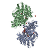
| ||||||||
|---|---|---|---|---|---|---|---|---|---|
| 1 | 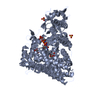
| ||||||||
| 2 | 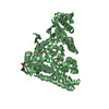
| ||||||||
| Unit cell |
|
-Components
-Protein , 1 types, 2 molecules AB
| #1: Protein | Mass: 80488.797 Da / Num. of mol.: 2 Source method: isolated from a genetically manipulated source Source: (gene. exp.) Mycobacterium tuberculosis (bacteria) / Gene: GlcB / Plasmid: pET15b / Species (production host): Escherichia coli / Production host: Escherichia coli BL21(DE3) (bacteria) / Strain (production host): BL21(DE3)References: UniProt: P0A5J4, UniProt: P9WK17*PLUS, EC: 4.1.3.2 |
|---|
-Non-polymers , 6 types, 649 molecules 


| #2: Chemical |  Mass: 24.305 Da / Num. of mol.: 2 / Source method: obtained synthetically / Formula: Mg Mass: 24.305 Da / Num. of mol.: 2 / Source method: obtained synthetically / Formula: Mg#3: Chemical | Sulfate Mass: 96.063 Da / Num. of mol.: 3 / Source method: obtained synthetically / Formula: SO4 Mass: 96.063 Da / Num. of mol.: 3 / Source method: obtained synthetically / Formula: SO4#4: Chemical | Malic acid Mass: 134.087 Da / Num. of mol.: 3 / Source method: obtained synthetically / Formula: C4H6O5 Mass: 134.087 Da / Num. of mol.: 3 / Source method: obtained synthetically / Formula: C4H6O5#5: Chemical | ChemComp-COA / | Coenzyme A Mass: 767.534 Da / Num. of mol.: 1 / Source method: obtained synthetically / Formula: C21H36N7O16P3S Mass: 767.534 Da / Num. of mol.: 1 / Source method: obtained synthetically / Formula: C21H36N7O16P3S#6: Chemical | ChemComp-GLV / | Glyoxylic acid Mass: 74.035 Da / Num. of mol.: 1 / Source method: obtained synthetically / Formula: C2H2O3 Mass: 74.035 Da / Num. of mol.: 1 / Source method: obtained synthetically / Formula: C2H2O3#7: Water | ChemComp-HOH / | WaterMass: 18.015 Da / Num. of mol.: 639 / Source method: isolated from a natural source / Formula: H2O |
|---|
-Experimental details
-Experiment
| Experiment | Method: X-RAY DIFFRACTION / Number of used crystals: 1 |
|---|
- Sample preparation
| Crystal | Density Matthews: 2.64 Å3/Da / Density % sol: 53.49 % | ||||||||||||||||||||||||||||||||||||||||||||||||
|---|---|---|---|---|---|---|---|---|---|---|---|---|---|---|---|---|---|---|---|---|---|---|---|---|---|---|---|---|---|---|---|---|---|---|---|---|---|---|---|---|---|---|---|---|---|---|---|---|---|
| Crystal grow | Temperature: 292 K / Method: vapor diffusion, hanging drop / pH: 6.5 Details: Ammonium Sulphate, MES pH 6.5, Dioxane, magnesium chloride, acetyl coenzyme A, glyoxylate, VAPOR DIFFUSION, HANGING DROP, temperature 292K | ||||||||||||||||||||||||||||||||||||||||||||||||
| Crystal grow | *PLUS pH: 8 | ||||||||||||||||||||||||||||||||||||||||||||||||
| Components of the solutions | *PLUS
|
-Data collection
| Diffraction | Mean temperature: 100 K |
|---|---|
| Diffraction source | Source: SYNCHROTRON / Site: APS  / Beamline: 14-BM-C / Wavelength: 1 Å / Beamline: 14-BM-C / Wavelength: 1 Å |
| Detector | Type: ADSC QUANTUM 4 / Detector: CCD / Date: Dec 15, 2000 / Details: mirrors |
| Radiation | Monochromator: bend cylindrical Ge(III) monochromator / Protocol: SINGLE WAVELENGTH / Monochromatic (M) / Laue (L): M / Scattering type: x-ray |
| Radiation wavelength | Wavelength: 1 Å / Relative weight: 1 |
| Reflection | Resolution: 2.6→30 Å / Num. obs: 51936 / % possible obs: 96.2 % / Observed criterion σ(F): 0 / Observed criterion σ(I): 0 |
| Reflection shell | Resolution: 2.6→2.69 Å / % possible all: 93.9 |
| Reflection | *PLUS Highest resolution: 2.7 Å / Lowest resolution: 30 Å / Num. measured all: 397417 / Rmerge(I) obs: 0.057 |
| Reflection shell | *PLUS % possible obs: 95.2 % / Rmerge(I) obs: 0.33 |
- Processing
| Software |
| ||||||||||||||||||||||||||||||||||||||||||||||||||||||||||||||||||||||
|---|---|---|---|---|---|---|---|---|---|---|---|---|---|---|---|---|---|---|---|---|---|---|---|---|---|---|---|---|---|---|---|---|---|---|---|---|---|---|---|---|---|---|---|---|---|---|---|---|---|---|---|---|---|---|---|---|---|---|---|---|---|---|---|---|---|---|---|---|---|---|---|
| Refinement | Method to determine structure: MOLECULAR REPLACEMENT / Resolution: 2.7→29.36 Å / Cor.coef. Fo:Fc: 0.939 / Cor.coef. Fo:Fc free: 0.858 / SU B: 17.822 / SU ML: 0.349 / Cross valid method: THROUGHOUT / σ(F): 0 / ESU R Free: 0.439 / Stereochemistry target values: MAXIMUM LIKELIHOOD
| ||||||||||||||||||||||||||||||||||||||||||||||||||||||||||||||||||||||
| Solvent computation | Ion probe radii: 0.8 Å / Shrinkage radii: 0.8 Å / VDW probe radii: 1.4 Å / Solvent model: BABINET MODEL WITH MASK | ||||||||||||||||||||||||||||||||||||||||||||||||||||||||||||||||||||||
| Displacement parameters | Biso mean: 25.996 Å2
| ||||||||||||||||||||||||||||||||||||||||||||||||||||||||||||||||||||||
| Refinement step | Cycle: LAST / Resolution: 2.7→29.36 Å
| ||||||||||||||||||||||||||||||||||||||||||||||||||||||||||||||||||||||
| Refine LS restraints |
| ||||||||||||||||||||||||||||||||||||||||||||||||||||||||||||||||||||||
| LS refinement shell | Resolution: 2.701→2.771 Å / Num. reflection Rfree: 308 / Num. reflection Rwork: 2772 / Total num. of bins used: 20 | ||||||||||||||||||||||||||||||||||||||||||||||||||||||||||||||||||||||
| Refinement | *PLUS Highest resolution: 2.7 Å / Lowest resolution: 30 Å / % reflection Rfree: 10 % / Rfactor Rfree: 0.287 / Rfactor Rwork: 0.19 | ||||||||||||||||||||||||||||||||||||||||||||||||||||||||||||||||||||||
| Solvent computation | *PLUS | ||||||||||||||||||||||||||||||||||||||||||||||||||||||||||||||||||||||
| Displacement parameters | *PLUS | ||||||||||||||||||||||||||||||||||||||||||||||||||||||||||||||||||||||
| Refine LS restraints | *PLUS
|
