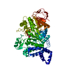
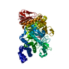
 Movie
Movie Controller


+ Open data

- Basic information
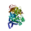
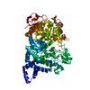
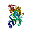
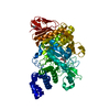
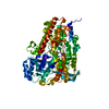
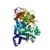
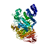
| Entry | Database: PDB / ID: 1mvy | |||||||||
|---|---|---|---|---|---|---|---|---|---|---|
| Title | Amylosucrase mutant E328Q co-crystallized with maltoheptaose. | |||||||||
 Components Components | amylosucrase | |||||||||
 Keywords Keywords | TRANSFERASE / (beta-alpha)8 barrel / protein-sugar complex | |||||||||
| Function / homology |  Function and homology informationamylosucrase / amylosucrase activity / carbohydrate metabolic process / extracellular region Function and homology informationamylosucrase / amylosucrase activity / carbohydrate metabolic process / extracellular regionSimilarity search - Function | |||||||||
| Biological species |  Neisseria polysaccharea (bacteria) Neisseria polysaccharea (bacteria) | |||||||||
| Method | X-RAY DIFFRACTION / SYNCHROTRON / MOLECULAR REPLACEMENT / Resolution: 2 Å | |||||||||
 Authors Authors | Skov, L.K. / Mirza, O. / Sprogoe, D. / Dar, I. / Remaud-Simeon, M. / Albenne, C. / Monsan, P. / Gajhede, M. | |||||||||
 Citation Citation | Journal: J.BIOL.CHEM. / Year: 2002 Title: Oligosaccharide and Sucrose Complexes of Amylosucrase. STRUCTURAL IMPLICATIONS FOR THE POLYMERASE ACTIVITY Authors: Skov, L.K. / Mirza, O. / Sprogoe, D. / Dar, I. / Remaud-Simeon, M. / Albenne, C. / Monsan, P. / Gajhede, M. #1: Journal: J.Biol.Chem. / Year: 2001Title: Amylosucrase, a Glucan-Synthesizing Enzyme from the Alpha-Amylase Family Authors: Skov, L.K. / Mirza, O. / Henriksen, A. / De Montalk, G.P. / Remaud-Simeon, M. / Sarcabal, P. / Willemot, R.M. / Monsan, P. / Gajhede, M. #2: Journal: Biochemistry / Year: 2001Title: Crystal Structure of Amylosucrase from Neisseria Polysaccharea in Complex with D-Glucose and the Active Site Mutant Glu328Gln in Complex with the Natural Substrate Sucrose Authors: Mirza, O. / Skov, L.K. / Remaud-Simeon, M. / De Montalk, G.P. / Albenne, C. / Monsan, P. / Gajhede, M. | |||||||||
| History |
|
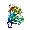
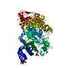
- Structure visualization
| Structure viewer | Molecule: MolmilJmol/JSmol |
|---|
- Downloads & links
-Download
| PDBx/mmCIF format | 1mvy.cif.gz | 148.1 KB | Display | PDBx/mmCIF format |
|---|---|---|---|---|
| PDB format | pdb1mvy.ent.gz | 114.7 KB | Display | PDB format |
| PDBx/mmJSON format | 1mvy.json.gz | Tree view | PDBx/mmJSON format | |
| Others |  Other downloads Other downloads |
-Validation report
| Arichive directory | https://data.pdbj.org/pub/pdb/validation_reports/mv/1mvyftp://data.pdbj.org/pub/pdb/validation_reports/mv/1mvy | HTTPS FTP |
|---|
-Related structure data


-Links
 PDBj
PDBj


- Assembly
| Deposited unit | 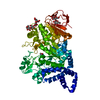
| ||||||||
|---|---|---|---|---|---|---|---|---|---|
| 1 |
| ||||||||
| Unit cell |
|
-Components
| #1: Protein | Mass: 71530.086 Da / Num. of mol.: 1 / Mutation: E328Q Source method: isolated from a genetically manipulated source Source: (gene. exp.) Neisseria polysaccharea (bacteria) / Production host: Escherichia coli (E. coli)References: GenBank: 4107260, UniProt: Q9ZEU2*PLUS, amylosucrase |
|---|---|
| #2: Polysaccharide | alpha-D-glucopyranose-(1-4)-alpha-D-glucopyranose-(1-4)-alpha-D-glucopyranose-(1-4)-alpha-D-glucopyranose / alpha-maltotetraose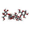  , Oligosaccharide / Class: Substrate analog / Mass: 666.578 Da / Num. of mol.: 1 , Oligosaccharide / Class: Substrate analog / Mass: 666.578 Da / Num. of mol.: 1Source method: isolated from a genetically manipulated source Details: oligosaccharide / References: alpha-maltotetraose |
| #3: Polysaccharide | alpha-D-glucopyranose-(1-4)-alpha-D-glucopyranose / alpha-maltose  , Oligosaccharide / Class: Nutrient / Mass: 342.297 Da / Num. of mol.: 1 , Oligosaccharide / Class: Nutrient / Mass: 342.297 Da / Num. of mol.: 1Source method: isolated from a genetically manipulated source Details: oligosaccharide / References: alpha-maltose |
| #4: Chemical | ChemComp-TRS / Tris  Mass: 122.143 Da / Num. of mol.: 1 / Source method: obtained synthetically / Formula: C4H12NO3 / Comment: pH buffer*YM Mass: 122.143 Da / Num. of mol.: 1 / Source method: obtained synthetically / Formula: C4H12NO3 / Comment: pH buffer*YM |
| #5: Water | ChemComp-HOH / Water Mass: 18.015 Da / Num. of mol.: 465 / Source method: isolated from a natural source / Formula: H2O Mass: 18.015 Da / Num. of mol.: 465 / Source method: isolated from a natural source / Formula: H2O |
-Experimental details
-Experiment
| Experiment | Method: X-RAY DIFFRACTION / Number of used crystals: 1 |
|---|
- Sample preparation
| Crystal | Density Matthews: 2.35 Å3/Da / Density % sol: 47.75 % | ||||||||||||||||||||||||||||||||||||||||||||||||||||||||
|---|---|---|---|---|---|---|---|---|---|---|---|---|---|---|---|---|---|---|---|---|---|---|---|---|---|---|---|---|---|---|---|---|---|---|---|---|---|---|---|---|---|---|---|---|---|---|---|---|---|---|---|---|---|---|---|---|---|
| Crystal grow | Temperature: 277 K / Method: vapor diffusion, hanging drop / pH: 7 Details: PEG 6000, SODIUM CHLORIDE, TRIS-HCL, EDTA, HEPES, DTT, maltoheptaose, pH 7, VAPOR DIFFUSION, HANGING DROP, temperature 277K | ||||||||||||||||||||||||||||||||||||||||||||||||||||||||
| Crystal grow | *PLUS pH: 7 / Details: Skov, L.K., (2000) Acta Crystallogr., D56, 203. | ||||||||||||||||||||||||||||||||||||||||||||||||||||||||
| Components of the solutions | *PLUS
|
-Data collection
| Diffraction | Mean temperature: 120 K |
|---|---|
| Diffraction source | Source: SYNCHROTRON / Site: MAX II  / Beamline: I711 / Wavelength: 1.016 Å / Beamline: I711 / Wavelength: 1.016 Å |
| Detector | Type: MARRESEARCH / Detector: CCD / Date: Mar 9, 2000 |
| Radiation | Monochromator: Si 111 CHANNEL / Protocol: SINGLE WAVELENGTH / Monochromatic (M) / Laue (L): M / Scattering type: x-ray |
| Radiation wavelength | Wavelength: 1.016 Å / Relative weight: 1 |
| Reflection | Resolution: 2→25 Å / Num. all: 45487 / Num. obs: 45487 / % possible obs: 97.5 % / Observed criterion σ(F): 0 / Observed criterion σ(I): 0 / Biso Wilson estimate: 8.8 Å2 / Rsym value: 0.062 |
| Reflection shell | Resolution: 2→2.07 Å / Rsym value: 0.22 / % possible all: 95.9 |
| Reflection | *PLUS Highest resolution: 2 Å / Lowest resolution: 25 Å / % possible obs: 97.9 % / Num. measured all: 413622 / Rmerge(I) obs: 0.062 |
| Reflection shell | *PLUS % possible obs: 95.5 % / Rmerge(I) obs: 0.22 |
- Processing
| Software |
| ||||||||||||||||||||||||||||||||||||
|---|---|---|---|---|---|---|---|---|---|---|---|---|---|---|---|---|---|---|---|---|---|---|---|---|---|---|---|---|---|---|---|---|---|---|---|---|---|
| Refinement | Method to determine structure: MOLECULAR REPLACEMENT / Resolution: 2→22.31 Å / Rfactor Rfree error: 0.005 / Isotropic thermal model: RESTRAINED / Cross valid method: THROUGHOUT / σ(F): 0 / Stereochemistry target values: Engh & Huber
| ||||||||||||||||||||||||||||||||||||
| Solvent computation | Solvent model: FLAT MODEL / Bsol: 38.2807 Å2 / ksol: 0.340482 e/Å3 | ||||||||||||||||||||||||||||||||||||
| Displacement parameters | Biso mean: 16.8 Å2
| ||||||||||||||||||||||||||||||||||||
| Refine analyze |
| ||||||||||||||||||||||||||||||||||||
| Refinement step | Cycle: LAST / Resolution: 2→22.31 Å
| ||||||||||||||||||||||||||||||||||||
| Refine LS restraints |
| ||||||||||||||||||||||||||||||||||||
| LS refinement shell | Resolution: 2→2.13 Å / Rfactor Rfree error: 0.014 / Total num. of bins used: 6
| ||||||||||||||||||||||||||||||||||||
| Xplor file |
| ||||||||||||||||||||||||||||||||||||
| Refinement | *PLUS Highest resolution: 2 Å / % reflection Rfree: 5 % | ||||||||||||||||||||||||||||||||||||
| Solvent computation | *PLUS | ||||||||||||||||||||||||||||||||||||
| Displacement parameters | *PLUS | ||||||||||||||||||||||||||||||||||||
| Refine LS restraints | *PLUS
|
