 Movie
Movie Controller

[English] 日本語
 Yorodumi
- PDB-1mvk: X-ray structure of the tetrameric mutant of the B1 domain of stre... -
+ Open data

- Basic information
| Entry | Database: PDB / ID: 1mvk | ||||||
|---|---|---|---|---|---|---|---|
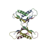
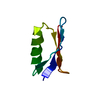
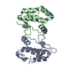
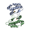
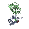
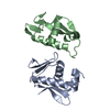
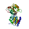
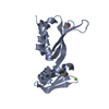
| Title | X-ray structure of the tetrameric mutant of the B1 domain of streptococcal protein G | ||||||
 Components Components | Immunoglobulin G binding protein G | ||||||
 Keywords Keywords | PROTEIN BINDING / strand-exchanged tetramer / channel | ||||||
| Function / homology |  Function and homology information Function and homology information | ||||||
| Biological species |  Streptococcus sp. 'group G' (bacteria) Streptococcus sp. 'group G' (bacteria) | ||||||
| Method |  X-RAY DIFFRACTION / MAD / Resolution: 2.5 Å X-RAY DIFFRACTION / MAD / Resolution: 2.5 Å | ||||||
 Authors Authors | Frank, M.K. / Dyda, F. / Dobrodumov, A. / Gronenborn, A.M. | ||||||
 Citation Citation | Journal: Nat.Struct.Biol. / Year: 2002 Title: Core mutations switch monomeric protein GB1 into an intertwined tetramer. Authors: Kirsten Frank, M. / Dyda, F. / Dobrodumov, A. / Gronenborn, A.M. #1: Journal: Science / Year: 1991Title: A Novel, Highly Stable Fold of the Immunoglobulin Binding Domain of Streptococcal Protein G Authors: Gronenborn, A.M. / Filpula, D.R. / Essig, N.Z. / Achari, A. / Whitlow, M. / Wingfield, P.T. / Clore, G.M. #2: Journal: FEBS Lett. / Year: 1996Title: Core Mutants of the Immunoglobulin Binding Domain of Streptococcal Protein G: Stability and Structural Integrity Authors: Gronenborn, A.M. / Frank, M.K. / Clore, G.M. | ||||||
| History |
|
- Structure visualization
| Structure viewer | Molecule: MolmilJmol/JSmol |
|---|
- Downloads & links
-Download
| PDBx/mmCIF format | 1mvk.cif.gz | 125.5 KB | Display | PDBx/mmCIF format |
|---|---|---|---|---|
| PDB format | pdb1mvk.ent.gz | 100.2 KB | Display | PDB format |
| PDBx/mmJSON format | 1mvk.json.gz | Tree view | PDBx/mmJSON format | |
| Others |  Other downloads Other downloads |
-Validation report
| Summary document | 1mvk_validation.pdf.gz | 450.1 KB | Display | wwPDB validaton report |
|---|---|---|---|---|
| Full document | 1mvk_full_validation.pdf.gz | 456.9 KB | Display | |
| Data in XML | 1mvk_validation.xml.gz | 11.4 KB | Display | |
| Data in CIF | 1mvk_validation.cif.gz | 19.9 KB | Display | |
| Arichive directory | https://data.pdbj.org/pub/pdb/validation_reports/mv/1mvkftp://data.pdbj.org/pub/pdb/validation_reports/mv/1mvk | HTTPS FTP |
-Related structure data


-Links
 PDBj
PDBj

- Assembly
| Deposited unit | 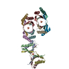
| ||||||||
|---|---|---|---|---|---|---|---|---|---|
| 1 | 
| ||||||||
| 2 | 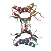
| ||||||||
| 3 | 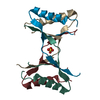
| ||||||||
| Unit cell |
| ||||||||
| Details | The asymmetric unit contains three copies of the biological unit. |
-Components
| #1: Antibody | Mass: 6302.931 Da / Num. of mol.: 12 / Fragment: B1 domain, sequence database residues 228-282 / Mutation: T2Q, L5V, A26F, F30V, Y33F, A34F Source method: isolated from a genetically manipulated source Source: (gene. exp.) Streptococcus sp. 'group G' (bacteria) / Gene: SPG / Plasmid: pET11a / Production host: #2: Chemical |   Mass: 96.063 Da / Num. of mol.: 3 / Source method: obtained synthetically / Formula: SO4 Mass: 96.063 Da / Num. of mol.: 3 / Source method: obtained synthetically / Formula: SO4#3: Water | ChemComp-HOH / |  Mass: 18.015 Da / Num. of mol.: 218 / Source method: isolated from a natural source / Formula: H2O Mass: 18.015 Da / Num. of mol.: 218 / Source method: isolated from a natural source / Formula: H2O |
|---|
-Experimental details
-Experiment
| Experiment | Method: X-RAY DIFFRACTION / Number of used crystals: 1 |
|---|
- Sample preparation
| Crystal | Density Matthews: 2.93 Å3/Da / Density % sol: 57.97 % | ||||||||||||||||||||||||||||||
|---|---|---|---|---|---|---|---|---|---|---|---|---|---|---|---|---|---|---|---|---|---|---|---|---|---|---|---|---|---|---|---|
| Crystal grow | Temperature: 293 K / Method: vapor diffusion, hanging drop / pH: 5.6 Details: PEG 8000, ammonium sulfate, sodium acetate, sodium chloride, TrisHCl, pH 5.6, VAPOR DIFFUSION, HANGING DROP, temperature 293K | ||||||||||||||||||||||||||||||
| Crystal grow | *PLUS | ||||||||||||||||||||||||||||||
| Components of the solutions | *PLUS
|
-Data collection
| Diffraction | Mean temperature: 95 K |
|---|---|
| Diffraction source | Source: ROTATING ANODE / Type: RIGAKU RU200 / Wavelength: 1.5418 Å |
| Detector | Type: RIGAKU RAXIS II / Detector: IMAGE PLATE / Date: Oct 7, 2000 / Details: total-reflection mirror pair |
| Radiation | Protocol: SINGLE WAVELENGTH / Monochromatic (M) / Laue (L): M / Scattering type: x-ray |
| Radiation wavelength | Wavelength: 1.5418 Å / Relative weight: 1 |
| Reflection | Resolution: 2.5→30 Å / Num. obs: 31523 / % possible obs: 99.4 % / Observed criterion σ(I): -3 / Redundancy: 5.78 % / Biso Wilson estimate: 28.35 Å2 / Rsym value: 0.083 / Net I/σ(I): 11.1 |
| Reflection shell | Resolution: 2.5→2.57 Å / % possible all: 93.4 |
| Reflection | *PLUS Num. measured all: 182446 / Rmerge(I) obs: 0.083 |
- Processing
| Software |
| ||||||||||||||||||||
|---|---|---|---|---|---|---|---|---|---|---|---|---|---|---|---|---|---|---|---|---|---|
| Refinement | Method to determine structure: MAD / Resolution: 2.5→30 Å / Cross valid method: THROUGHOUT / σ(F): 0 / Stereochemistry target values: Engh & Huber Details: Flexible region from residues 8-21 missing in electron density of most chains
| ||||||||||||||||||||
| Refinement step | Cycle: LAST / Resolution: 2.5→30 Å
| ||||||||||||||||||||
| Refine LS restraints |
| ||||||||||||||||||||
| LS refinement shell | Resolution: 2.5→2.61 Å / Rfactor Rfree error: 0.03088
| ||||||||||||||||||||
| Refinement | *PLUS Lowest resolution: 30 Å / % reflection Rfree: 5 % / Rfactor obs: 0.237 / Rfactor Rfree: 0.283 / Rfactor Rwork: 0.237 | ||||||||||||||||||||
| Solvent computation | *PLUS | ||||||||||||||||||||
| Displacement parameters | *PLUS | ||||||||||||||||||||
| Refine LS restraints | *PLUS
| ||||||||||||||||||||
| LS refinement shell | *PLUS Rfactor Rfree: 0.3882 / Rfactor Rwork: 0.3912 |
