 Movie
Movie Controller

[English] 日本語
 Yorodumi
- PDB-1mto: Crystal structure of a Phosphofructokinase mutant from Bacillus s... -
+ Open data

- Basic information
| Entry | Database: PDB / ID: 1mto | ||||||
|---|---|---|---|---|---|---|---|
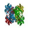
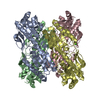
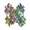
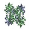
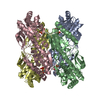
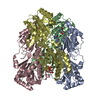
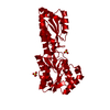
| Title | Crystal structure of a Phosphofructokinase mutant from Bacillus stearothermophilus bound with fructose-6-phosphate | ||||||
 Components Components | 6-phosphofructokinase Phosphofructokinase 1 Phosphofructokinase 1 | ||||||
 Keywords Keywords | TRANSFERASE / phosphofructokinase / fructose-6-phosphate / tryptophan-shift | ||||||
| Function / homology |  Function and homology information6-phosphofructokinase / 6-phosphofructokinase activity / fructose 6-phosphate metabolic process / ATP binding / metal ion binding / cytoplasm Function and homology information6-phosphofructokinase / 6-phosphofructokinase activity / fructose 6-phosphate metabolic process / ATP binding / metal ion binding / cytoplasmSimilarity search - Function | ||||||
| Biological species |   Geobacillus stearothermophilus (bacteria) Geobacillus stearothermophilus (bacteria) | ||||||
| Method | X-RAY DIFFRACTION / MOLECULAR REPLACEMENT / Resolution: 3.2 Å | ||||||
 Authors Authors | Riley-Lovingshimer, M.R. / Ronning, D.R. / Sacchettini, J.C. / Reinhart, G.D. | ||||||
 Citation Citation | Journal: Biochemistry / Year: 2002 Title: Reversible Ligand-Induced Dissociation of a Tryptophan-Shift Mutant of Phosphofructokinase from Bacillus stearothermophilus Authors: Riley-Lovingshimer, M.R. / Ronning, D.R. / Sacchettini, J.C. / Reinhart, G.D. | ||||||
| History |
|
- Structure visualization
| Structure viewer | Molecule: MolmilJmol/JSmol |
|---|
- Downloads & links
-Download
| PDBx/mmCIF format | 1mto.cif.gz | 466.5 KB | Display | PDBx/mmCIF format |
|---|---|---|---|---|
| PDB format | pdb1mto.ent.gz | 388.3 KB | Display | PDB format |
| PDBx/mmJSON format | 1mto.json.gz | Tree view | PDBx/mmJSON format | |
| Others |  Other downloads Other downloads |
-Validation report
| Arichive directory | https://data.pdbj.org/pub/pdb/validation_reports/mt/1mtoftp://data.pdbj.org/pub/pdb/validation_reports/mt/1mto | HTTPS FTP |
|---|
-Related structure data
| Related structure data |  3pfkS S: Starting model for refinement |
|---|---|
| Similar structure data |

-Links
 PDBj
PDBj
- Assembly
| Deposited unit | 
| ||||||||
|---|---|---|---|---|---|---|---|---|---|
| 1 | 
| ||||||||
| 2 | 
| ||||||||
| Unit cell |
|
-Components
| #1: Protein | Phosphofructokinase 1 / Phosphofructokinase / Phosphohexokinase Mass: 34167.852 Da / Num. of mol.: 8 / Mutation: W179Y,Y164W Source method: isolated from a genetically manipulated source Source: (gene. exp.) Geobacillus stearothermophilus (bacteria)Gene: pfk / Production host: Escherichia coli (E. coli) / Strain (production host): DF1020 / References: UniProt: P00512, 6-phosphofructokinase#2: Sugar | ChemComp-F6P / Fructose 6-phosphate  Type: D-saccharide, beta linking / Mass: 260.136 Da / Num. of mol.: 8 Type: D-saccharide, beta linking / Mass: 260.136 Da / Num. of mol.: 8Source method: isolated from a genetically manipulated source Formula: C6H13O9P |
|---|
-Experimental details
-Experiment
| Experiment | Method: X-RAY DIFFRACTION / Number of used crystals: 1 |
|---|
- Sample preparation
| Crystal | Density Matthews: 2.368 Å3/Da / Density % sol: 44.5707 % | ||||||||||||||||||||||||||||||||||||
|---|---|---|---|---|---|---|---|---|---|---|---|---|---|---|---|---|---|---|---|---|---|---|---|---|---|---|---|---|---|---|---|---|---|---|---|---|---|
| Crystal grow | Temperature: 291 K / Method: vapor diffusion, hanging drop / pH: 6.5 Details: PEG 8000, Sodium Cacodylate, Calcium Acetate, Fructose-6-phosphate, pH 6.5, VAPOR DIFFUSION, HANGING DROP, temperature 291K | ||||||||||||||||||||||||||||||||||||
| Crystal grow | *PLUS | ||||||||||||||||||||||||||||||||||||
| Components of the solutions | *PLUS
|
-Data collection
| Diffraction | Mean temperature: 120 K |
|---|---|
| Diffraction source | Source: ROTATING ANODE / Type: RIGAKU RU200 / Wavelength: 1.5418 Å |
| Detector | Type: MACSCIENCE / Detector: IMAGE PLATE / Date: Mar 5, 2000 |
| Radiation | Protocol: SINGLE WAVELENGTH / Monochromatic (M) / Laue (L): M / Scattering type: x-ray |
| Radiation wavelength | Wavelength: 1.5418 Å / Relative weight: 1 |
| Reflection | Resolution: 3.2→29.84 Å / Num. obs: 46829 / Observed criterion σ(F): 0 |
| Reflection | *PLUS Highest resolution: 3.1 Å / % possible obs: 91.6 % / Num. measured all: 106908 / Rmerge(I) obs: 0.101 |
| Reflection shell | *PLUS % possible obs: 89.7 % |
- Processing
| Software |
| ||||||||||||||||||||||||||||||||||||||||||||||||||||||||||||||||||||||||||||||||
|---|---|---|---|---|---|---|---|---|---|---|---|---|---|---|---|---|---|---|---|---|---|---|---|---|---|---|---|---|---|---|---|---|---|---|---|---|---|---|---|---|---|---|---|---|---|---|---|---|---|---|---|---|---|---|---|---|---|---|---|---|---|---|---|---|---|---|---|---|---|---|---|---|---|---|---|---|---|---|---|---|---|
| Refinement | Method to determine structure: MOLECULAR REPLACEMENT Starting model: PDB ENTRY 3PFK Resolution: 3.2→29.84 Å / Rfactor Rfree error: 0.005 / Data cutoff high absF: 190041.81 / Data cutoff high rms absF: 190041.81 / Data cutoff low absF: 0 / Isotropic thermal model: RESTRAINED / Cross valid method: THROUGHOUT / σ(F): 0
| ||||||||||||||||||||||||||||||||||||||||||||||||||||||||||||||||||||||||||||||||
| Solvent computation | Solvent model: FLAT MODEL / Bsol: 10 Å2 / ksol: 0.250378 e/Å3 | ||||||||||||||||||||||||||||||||||||||||||||||||||||||||||||||||||||||||||||||||
| Displacement parameters | Biso mean: 31.8 Å2
| ||||||||||||||||||||||||||||||||||||||||||||||||||||||||||||||||||||||||||||||||
| Refine analyze |
| ||||||||||||||||||||||||||||||||||||||||||||||||||||||||||||||||||||||||||||||||
| Refinement step | Cycle: LAST / Resolution: 3.2→29.84 Å
| ||||||||||||||||||||||||||||||||||||||||||||||||||||||||||||||||||||||||||||||||
| Refine LS restraints |
| ||||||||||||||||||||||||||||||||||||||||||||||||||||||||||||||||||||||||||||||||
| LS refinement shell | Resolution: 3.2→3.4 Å / Rfactor Rfree error: 0.015 / Total num. of bins used: 6
| ||||||||||||||||||||||||||||||||||||||||||||||||||||||||||||||||||||||||||||||||
| Xplor file |
| ||||||||||||||||||||||||||||||||||||||||||||||||||||||||||||||||||||||||||||||||
| Refinement | *PLUS Highest resolution: 3.1 Å / Num. reflection obs: 32894 / Rfactor Rfree: 0.2746 / Rfactor Rwork: 0.1787 | ||||||||||||||||||||||||||||||||||||||||||||||||||||||||||||||||||||||||||||||||
| Solvent computation | *PLUS | ||||||||||||||||||||||||||||||||||||||||||||||||||||||||||||||||||||||||||||||||
| Displacement parameters | *PLUS | ||||||||||||||||||||||||||||||||||||||||||||||||||||||||||||||||||||||||||||||||
| Refine LS restraints | *PLUS
|
