 Movie
Movie Controller


+ Open data

- Basic information
| Entry | Database: PDB / ID: 1mq0 | ||||||
|---|---|---|---|---|---|---|---|
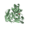
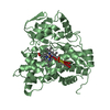
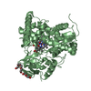
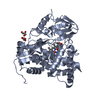
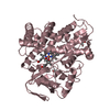
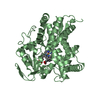
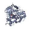
| Title | Crystal Structure of Human Cytidine Deaminase | ||||||
 Components Components | Cytidine Deaminase | ||||||
 Keywords Keywords | HYDROLASE / human / enzyme / cytidine deaminase / amine hydrolase / inhibitor / diazepinone / leukemia / chemotherapy / anticancer / drug / phi-phi interaction / edge-to-face interaction / protein | ||||||
| Function / homology |  Function and homology information Function and homology informationpyrimidine nucleoside salvage / negative regulation of nucleotide metabolic process / pyrimidine-containing compound salvage / cytidine deaminase / cytidine deamination / neutrophil degranulation / Pyrimidine salvage / cytidine deaminase activity / nucleoside binding / cytosine metabolic process ...pyrimidine nucleoside salvage / negative regulation of nucleotide metabolic process / pyrimidine-containing compound salvage / cytidine deaminase / cytidine deamination / neutrophil degranulation / Pyrimidine salvage / cytidine deaminase activity / nucleoside binding / cytosine metabolic process / negative regulation of cell growth / tertiary granule lumen / secretory granule lumen / ficolin-1-rich granule lumen / cell surface receptor signaling pathway / Neutrophil degranulation / protein homodimerization activity / zinc ion binding / extracellular region / identical protein binding / cytosolSimilarity search - Function | ||||||
| Biological species |  Homo sapiens (human) Homo sapiens (human) | ||||||
| Method | X-RAY DIFFRACTION / MOLECULAR REPLACEMENT / Resolution: 2.4 Å | ||||||
 Authors Authors | Chung, S.J. / Fromme, J.C. / Verdine, G.L. | ||||||
 Citation Citation | Journal: J.Med.Chem. / Year: 2005 Title: Structure of human cytidine deaminase bound to a potent inhibitor Authors: Chung, S.J. / Fromme, J.C. / Verdine, G.L. | ||||||
| History |
|
- Structure visualization
| Structure viewer | Molecule: MolmilJmol/JSmol |
|---|
- Downloads & links
-Download
| PDBx/mmCIF format | 1mq0.cif.gz | 64.7 KB | Display | PDBx/mmCIF format |
|---|---|---|---|---|
| PDB format | pdb1mq0.ent.gz | 46.7 KB | Display | PDB format |
| PDBx/mmJSON format | 1mq0.json.gz | Tree view | PDBx/mmJSON format | |
| Others |  Other downloads Other downloads |
-Validation report
| Arichive directory | https://data.pdbj.org/pub/pdb/validation_reports/mq/1mq0ftp://data.pdbj.org/pub/pdb/validation_reports/mq/1mq0 | HTTPS FTP |
|---|
-Related structure data
| Related structure data | 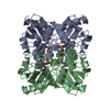 1jtkS S: Starting model for refinement |
|---|---|
| Similar structure data |


-Links
 PDBj
PDBj- Assembly
| Deposited unit | 
| ||||||||
|---|---|---|---|---|---|---|---|---|---|
| 1 | 
| ||||||||
| Unit cell |
| ||||||||
| Details | The biological unit is a tetramer, and there is a dimer in the asymmetric unit. The other dimer of the tetramer can be obtained by the following symmetry operation: (-X, -Y,Z) dx=1 dy=3 dz=0 |
-Components
| #1: Protein | / Cytidine aminohydrolase Mass: 15510.718 Da / Num. of mol.: 2 / Mutation: K27Q Source method: isolated from a genetically manipulated source Source: (gene. exp.) Homo sapiens (human) / Species (production host): Escherichia coli / Production host:  Escherichia coli BL21 (bacteria) / Strain (production host): BL21 / References: UniProt: P32320, cytidine deaminase Escherichia coli BL21 (bacteria) / Strain (production host): BL21 / References: UniProt: P32320, cytidine deaminase#2: Chemical |   Mass: 65.409 Da / Num. of mol.: 2 / Source method: obtained synthetically / Formula: Zn Mass: 65.409 Da / Num. of mol.: 2 / Source method: obtained synthetically / Formula: Zn#3: Chemical |   Mass: 242.229 Da / Num. of mol.: 2 / Source method: obtained synthetically / Formula: C10H14N2O5 Mass: 242.229 Da / Num. of mol.: 2 / Source method: obtained synthetically / Formula: C10H14N2O5#4: Water | ChemComp-HOH / | Water Mass: 18.015 Da / Num. of mol.: 62 / Source method: isolated from a natural source / Formula: H2O Mass: 18.015 Da / Num. of mol.: 62 / Source method: isolated from a natural source / Formula: H2O |
|---|
-Experimental details
-Experiment
| Experiment | Method: X-RAY DIFFRACTION / Number of used crystals: 1 |
|---|
- Sample preparation
| Crystal | Density Matthews: 2.13 Å3/Da / Density % sol: 42.32 % | |||||||||||||||||||||||||||||||||||||||||||||||||||||||||||||||
|---|---|---|---|---|---|---|---|---|---|---|---|---|---|---|---|---|---|---|---|---|---|---|---|---|---|---|---|---|---|---|---|---|---|---|---|---|---|---|---|---|---|---|---|---|---|---|---|---|---|---|---|---|---|---|---|---|---|---|---|---|---|---|---|---|
| Crystal grow | Temperature: 298 K / Method: vapor diffusion, hanging drop / pH: 4.6 Details: sodium acetate, 2-methyl-2,4-pentane-diol, calcium chloride, synthetic inhibitor, pH 4.6, VAPOR DIFFUSION, HANGING DROP, temperature 298K | |||||||||||||||||||||||||||||||||||||||||||||||||||||||||||||||
| Crystal grow | *PLUS Temperature: 4 ℃ / pH: 7.5 / Method: vapor diffusion, hanging drop | |||||||||||||||||||||||||||||||||||||||||||||||||||||||||||||||
| Components of the solutions | *PLUS
|
-Data collection
| Diffraction | Mean temperature: 100 K |
|---|---|
| Diffraction source | Source: ROTATING ANODE / Type: RIGAKU ULTRAX 18 / Wavelength: 1.5418 Å |
| Detector | Type: RIGAKU RAXIS IV / Detector: IMAGE PLATE / Date: Jul 19, 2002 / Details: Osmic Blue |
| Radiation | Monochromator: Nickel filter / Protocol: SINGLE WAVELENGTH / Monochromatic (M) / Laue (L): M / Scattering type: x-ray |
| Radiation wavelength | Wavelength: 1.5418 Å / Relative weight: 1 |
| Reflection | Resolution: 2.3→47.45 Å / Num. obs: 11082 / % possible obs: 97.1 % / Observed criterion σ(I): 0 / Redundancy: 4.58 % / Biso Wilson estimate: 28.5 Å2 / Rmerge(I) obs: 0.09 / Net I/σ(I): 10.4 |
| Reflection shell | Resolution: 2.3→2.38 Å / Redundancy: 4.56 % / Rmerge(I) obs: 0.326 / Mean I/σ(I) obs: 2.9 / Num. unique all: 1134 / % possible all: 98.1 |
| Reflection | *PLUS Highest resolution: 2.3 Å / Num. obs: 22165 / % possible obs: 97.1 % / Redundancy: 4.4 % |
| Reflection shell | *PLUS % possible obs: 98.1 % / Redundancy: 4.5 % / Rmerge(I) obs: 0.33 |
- Processing
| Software |
| ||||||||||||||||||||||||||||||||||||
|---|---|---|---|---|---|---|---|---|---|---|---|---|---|---|---|---|---|---|---|---|---|---|---|---|---|---|---|---|---|---|---|---|---|---|---|---|---|
| Refinement | Method to determine structure: MOLECULAR REPLACEMENT Starting model: PDB ENTRY 1JTK Resolution: 2.4→47.45 Å / Rfactor Rfree error: 0.011 / Data cutoff high absF: 210371.16 / Data cutoff high rms absF: 210371.16 / Data cutoff low absF: 0 / Isotropic thermal model: RESTRAINED / Cross valid method: THROUGHOUT / σ(F): 0 / Stereochemistry target values: Engh & Huber
| ||||||||||||||||||||||||||||||||||||
| Solvent computation | Solvent model: FLAT MODEL / Bsol: 51.7547 Å2 / ksol: 0.338176 e/Å3 | ||||||||||||||||||||||||||||||||||||
| Displacement parameters | Biso mean: 38.9 Å2
| ||||||||||||||||||||||||||||||||||||
| Refine analyze |
| ||||||||||||||||||||||||||||||||||||
| Refinement step | Cycle: LAST / Resolution: 2.4→47.45 Å
| ||||||||||||||||||||||||||||||||||||
| Refine LS restraints |
| ||||||||||||||||||||||||||||||||||||
| LS refinement shell | Resolution: 2.4→2.55 Å / Rfactor Rfree error: 0.037 / Total num. of bins used: 6
| ||||||||||||||||||||||||||||||||||||
| Xplor file |
| ||||||||||||||||||||||||||||||||||||
| Refinement | *PLUS % reflection Rfree: 10 % | ||||||||||||||||||||||||||||||||||||
| Solvent computation | *PLUS | ||||||||||||||||||||||||||||||||||||
| Displacement parameters | *PLUS | ||||||||||||||||||||||||||||||||||||
| Refine LS restraints | *PLUS
|
