 Movie
Movie Controller

[English] 日本語
 Yorodumi
- PDB-1m7u: Crystal structure of a novel DNA-binding domain from Ndt80, a tra... -
+ Open data

- Basic information
| Entry | Database: PDB / ID: 1m7u | ||||||
|---|---|---|---|---|---|---|---|
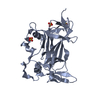
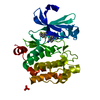
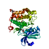
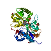
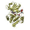
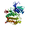
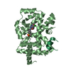
| Title | Crystal structure of a novel DNA-binding domain from Ndt80, a transcriptional activator required for meiosis in yeast | ||||||
 Components Components | Ndt80 protein | ||||||
 Keywords Keywords |  TRANSCRIPTION ACTIVATOR / yeast protein / DNA-binding / meiosis TRANSCRIPTION ACTIVATOR / yeast protein / DNA-binding / meiosis | ||||||
| Function / homology |  Function and homology information Function and homology informationnuclear chromosome / meiotic cell cycle / sequence-specific DNA binding / DNA-binding transcription factor activity / cell division / positive regulation of transcription by RNA polymerase IISimilarity search - Function | ||||||
| Biological species |  Saccharomyces cerevisiae (brewer's yeast) Saccharomyces cerevisiae (brewer's yeast) | ||||||
| Method | X-RAY DIFFRACTION / SYNCHROTRON / SIRAS / Resolution: 2.8 Å | ||||||
 Authors Authors | Montano, S.P. / Cote, M.L. / Fingerman, I. / Pierce, M. / Vershon, A.K. / Georgiadis, M.M. | ||||||
 Citation Citation | Journal: Proc.Natl.Acad.Sci.USA / Year: 2002 Title: Crystal structure of the DNA-binding domain from Ndt80, a transcriptional activator required for meiosis in yeast Authors: Montano, S.P. / Cote, M.L. / Fingerman, I. / Pierce, M. / Vershon, A.K. / Georgiadis, M.M. | ||||||
| History |
| ||||||
| Remark 999 | SEQUENCE THE DISCREPANCIES ARE PRESENT DUE TO ERRORS IN PCR. |
- Structure visualization
| Structure viewer | Molecule: MolmilJmol/JSmol |
|---|
- Downloads & links
-Download
| PDBx/mmCIF format | 1m7u.cif.gz | 106.7 KB | Display | PDBx/mmCIF format |
|---|---|---|---|---|
| PDB format | pdb1m7u.ent.gz | 81.9 KB | Display | PDB format |
| PDBx/mmJSON format | 1m7u.json.gz | Tree view | PDBx/mmJSON format | |
| Others |  Other downloads Other downloads |
-Validation report
| Arichive directory | https://data.pdbj.org/pub/pdb/validation_reports/m7/1m7uftp://data.pdbj.org/pub/pdb/validation_reports/m7/1m7u | HTTPS FTP |
|---|
-Related structure data


-Links
 PDBj
PDBj


- Assembly
| Deposited unit | 
| ||||||||
|---|---|---|---|---|---|---|---|---|---|
| 1 | 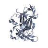
| ||||||||
| 2 | 
| ||||||||
| Unit cell |
|
-Components
| #1: Protein | Mass: 31307.455 Da / Num. of mol.: 2 / Fragment: DNA binding domain, residues 59-330 Source method: isolated from a genetically manipulated source Source: (gene. exp.) Saccharomyces cerevisiae (brewer's yeast)Gene: Ndt80 / Plasmid: pET15b / Production host:  Escherichia coli (E. coli) / Strain (production host): BL21 Codon+ / References: UniProt: P38830 Escherichia coli (E. coli) / Strain (production host): BL21 Codon+ / References: UniProt: P38830#2: Water | ChemComp-HOH / | Water Mass: 18.015 Da / Num. of mol.: 46 / Source method: isolated from a natural source / Formula: H2O Mass: 18.015 Da / Num. of mol.: 46 / Source method: isolated from a natural source / Formula: H2O |
|---|
-Experimental details
-Experiment
| Experiment | Method: X-RAY DIFFRACTION / Number of used crystals: 1 |
|---|
- Sample preparation
| Crystal | Density Matthews: 2.69 Å3/Da / Density % sol: 54.23 % | |||||||||||||||||||||||||||||||||||||||||||||||||
|---|---|---|---|---|---|---|---|---|---|---|---|---|---|---|---|---|---|---|---|---|---|---|---|---|---|---|---|---|---|---|---|---|---|---|---|---|---|---|---|---|---|---|---|---|---|---|---|---|---|---|
| Crystal grow | Temperature: 298 K / Method: vapor diffusion, hanging drop / pH: 5.6 Details: ammonium sulfate, citric acid, pH 5.6, VAPOR DIFFUSION, HANGING DROP, temperature 298K | |||||||||||||||||||||||||||||||||||||||||||||||||
| Crystal grow | *PLUS Temperature: 294 K | |||||||||||||||||||||||||||||||||||||||||||||||||
| Components of the solutions | *PLUS
|
-Data collection
| Diffraction | Mean temperature: 108 K |
|---|---|
| Diffraction source | Source: SYNCHROTRON / Site: NSLS  / Beamline: X25 / Wavelength: 1.1 Å / Beamline: X25 / Wavelength: 1.1 Å |
| Detector | Type: BRANDEIS - B4 / Detector: CCD / Date: Jun 29, 2001 |
| Radiation | Monochromator: Si 111 Channel / Protocol: SINGLE WAVELENGTH / Monochromatic (M) / Laue (L): M / Scattering type: x-ray |
| Radiation wavelength | Wavelength: 1.1 Å / Relative weight: 1 |
| Reflection | Resolution: 2.8→20 Å / Num. all: 17541 / Num. obs: 17150 / % possible obs: 97.8 % / Observed criterion σ(F): 2 / Observed criterion σ(I): 2 / Biso Wilson estimate: 39 Å2 / Rsym value: 0.067 / Net I/σ(I): 18.6 |
| Reflection shell | Resolution: 2.8→2.86 Å / Mean I/σ(I) obs: 6.8 / Rsym value: 0.21 / % possible all: 89.9 |
| Reflection | *PLUS % possible obs: 98.7 % / Rmerge(I) obs: 0.067 |
| Reflection shell | *PLUS Lowest resolution: 2.9 Å / % possible obs: 99.9 % / Rmerge(I) obs: 0.209 / Mean I/σ(I) obs: 6.8 |
- Processing
| Software |
| ||||||||||||||||||||
|---|---|---|---|---|---|---|---|---|---|---|---|---|---|---|---|---|---|---|---|---|---|
| Refinement | Method to determine structure: SIRAS / Resolution: 2.8→20 Å / σ(F): 0 / σ(I): 0 / Stereochemistry target values: Engh & Huber / Details: maximum likelihood
| ||||||||||||||||||||
| Refinement step | Cycle: LAST / Resolution: 2.8→20 Å
| ||||||||||||||||||||
| LS refinement shell | Resolution: 2.8→2.86 Å /
| ||||||||||||||||||||
| Refinement | *PLUS % reflection Rfree: 5 % / Rfactor Rfree: 0.29 / Rfactor Rwork: 0.232 | ||||||||||||||||||||
| Solvent computation | *PLUS | ||||||||||||||||||||
| Displacement parameters | *PLUS | ||||||||||||||||||||
| Refine LS restraints | *PLUS
| ||||||||||||||||||||
| LS refinement shell | *PLUS Rfactor Rfree: 0.37 / Rfactor Rwork: 0.32 |
