 Movie
Movie Controller

[English] 日本語
 Yorodumi
- PDB-2xiy: Protein kinase Pim-1 in complex with fragment-2 from crystallogra... -
+ Open data

- Basic information
| Entry | Database: PDB / ID: 2xiy | ||||||
|---|---|---|---|---|---|---|---|
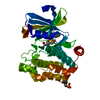
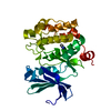
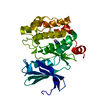
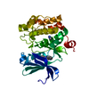
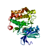
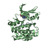
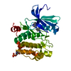
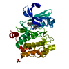
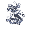
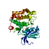
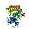
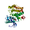
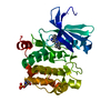
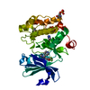
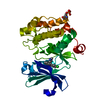
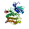
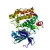
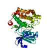
| Title | Protein kinase Pim-1 in complex with fragment-2 from crystallographic fragment screen | ||||||
 Components Components | PROTO-ONCOGENE SERINE/THREONINE PROTEIN KINASE PIM-1 | ||||||
 Keywords Keywords |  TRANSFERASE / PHOSPHORYLATION TRANSFERASE / PHOSPHORYLATION | ||||||
| Function / homology |  Function and homology information Function and homology informationpositive regulation of cardioblast proliferation / cellular detoxification / regulation of hematopoietic stem cell proliferation / vitamin D receptor signaling pathway / STAT5 activation downstream of FLT3 ITD mutants / transcription factor binding / ribosomal small subunit binding / positive regulation of cyclin-dependent protein serine/threonine kinase activity / positive regulation of cardiac muscle cell proliferation / positive regulation of TORC1 signaling ...positive regulation of cardioblast proliferation / cellular detoxification / regulation of hematopoietic stem cell proliferation / vitamin D receptor signaling pathway / STAT5 activation downstream of FLT3 ITD mutants / transcription factor binding / ribosomal small subunit binding / positive regulation of cyclin-dependent protein serine/threonine kinase activity / positive regulation of cardiac muscle cell proliferation / positive regulation of TORC1 signaling / Signaling by FLT3 fusion proteins / negative regulation of innate immune response / positive regulation of brown fat cell differentiation / protein serine/threonine kinase activator activity / regulation of transmembrane transporter activity / positive regulation of protein serine/threonine kinase activity / negative regulation of DNA-binding transcription factor activity / cellular response to type II interferon / manganese ion binding / Interleukin-4 and Interleukin-13 signaling / protein autophosphorylation / protein stabilization / non-specific serine/threonine protein kinase / cell cycle / protein phosphorylation / protein serine kinase activity / protein serine/threonine kinase activity / apoptotic process / nucleolus / negative regulation of apoptotic process / positive regulation of DNA-templated transcription / nucleoplasm / ATP binding / nucleus / plasma membrane / cytosol / cytoplasmSimilarity search - Function | ||||||
| Biological species |  HOMO SAPIENS (human) HOMO SAPIENS (human) | ||||||
| Method | X-RAY DIFFRACTION / SYNCHROTRON / MOLECULAR REPLACEMENT / Resolution: 2.2 Å | ||||||
 Authors Authors | Schulz, M.N. / Fanghanel, J. / Schafer, M. / Badock, V. / Briem, H. / Boemer, U. / Nguyen, D. / Husemann, M. / Hillig, R.C. | ||||||
 Citation Citation | Journal: Acta Crystallogr.,Sect.D / Year: 2011 Title: Crystallographic Fragment Screen Identifies Cinnamic Acid Derivatives as Starting Points for Potent Pim-1 Inhibitors Authors: Schulz, M.N. / Fanghanel, J. / Schafer, M. / Badock, V. / Briem, H. / Boemer, U. / Nguyen, D. / Husemann, M. / Hillig, R.C. | ||||||
| History |
|
- Structure visualization
| Structure viewer | Molecule: MolmilJmol/JSmol |
|---|
- Downloads & links
-Download
| PDBx/mmCIF format | 2xiy.cif.gz | 132.5 KB | Display | PDBx/mmCIF format |
|---|---|---|---|---|
| PDB format | pdb2xiy.ent.gz | 103 KB | Display | PDB format |
| PDBx/mmJSON format | 2xiy.json.gz | Tree view | PDBx/mmJSON format | |
| Others |  Other downloads Other downloads |
-Validation report
| Arichive directory | https://data.pdbj.org/pub/pdb/validation_reports/xi/2xiyftp://data.pdbj.org/pub/pdb/validation_reports/xi/2xiy | HTTPS FTP |
|---|
-Related structure data


-Links
 PDBj
PDBj

- Assembly
| Deposited unit | 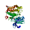
| ||||||||
|---|---|---|---|---|---|---|---|---|---|
| 1 |
| ||||||||
| Unit cell |
|
-Components
-Protein , 1 types, 1 molecules A
| #1: Protein | Mass: 34388.934 Da / Num. of mol.: 1 / Fragment: KINASE DOMAIN, RESIDUES 14-313 Source method: isolated from a genetically manipulated source Source: (gene. exp.) HOMO SAPIENS (human) / Plasmid: PET30A, HIS-TEV-PIM-1(14-313) / Production host:  ESCHERICHIA COLI (E. coli) ESCHERICHIA COLI (E. coli)References: UniProt: P11309, EC: 2.7.1.37, non-specific serine/threonine protein kinase |
|---|
-Non-polymers , 5 types, 136 molecules 


| #2: Chemical | ChemComp-XIY /  Mass: 148.162 Da / Num. of mol.: 1 / Source method: obtained synthetically / Formula: C8H8N2O Mass: 148.162 Da / Num. of mol.: 1 / Source method: obtained synthetically / Formula: C8H8N2O |
|---|---|
| #3: Chemical | ChemComp-DMS / Dimethyl sulfoxide Mass: 78.133 Da / Num. of mol.: 1 / Source method: obtained synthetically / Formula: C2H6OS / Comment: DMSO, precipitant*YM Mass: 78.133 Da / Num. of mol.: 1 / Source method: obtained synthetically / Formula: C2H6OS / Comment: DMSO, precipitant*YM |
| #4: Chemical | ChemComp-CL / Chloride Mass: 35.453 Da / Num. of mol.: 1 / Source method: obtained synthetically / Formula: Cl Mass: 35.453 Da / Num. of mol.: 1 / Source method: obtained synthetically / Formula: Cl |
| #5: Chemical | ChemComp-GOL / Glycerol Mass: 92.094 Da / Num. of mol.: 1 / Source method: obtained synthetically / Formula: C3H8O3 Mass: 92.094 Da / Num. of mol.: 1 / Source method: obtained synthetically / Formula: C3H8O3 |
| #6: Water | ChemComp-HOH / WaterMass: 18.015 Da / Num. of mol.: 132 / Source method: isolated from a natural source / Formula: H2O |
-Details
| Nonpolymer details | GLYCEROL (GOL): ARTIFACT FROM CRYO BUFFER CHLORIDE ION (CL): ARTIFACT FROM CRYSTALLIZATION BUFFER ...GLYCEROL (GOL): ARTIFACT FROM CRYO BUFFER CHLORIDE ION (CL): ARTIFACT FROM CRYSTALLIZ |
|---|---|
| Sequence details | N-TERMINAL GLY FROM TEV CLEAVAGE SITE |
-Experimental details
-Experiment
| Experiment | Method: X-RAY DIFFRACTION / Number of used crystals: 1 |
|---|
- Sample preparation
| Crystal | Density Matthews: 3.28 Å3/Da / Density % sol: 62.8 % / Description: SOLVED BY RIGID BODY REFINEMENT |
|---|---|
| Crystal grow | pH: 5.5 Details: 0.9 M (NH4)2HPO4,0.1 M SODIUM CITRATE PH=5.5 , 0.2M NACL |
-Data collection
| Diffraction | Mean temperature: 100 K |
|---|---|
| Diffraction source | Source: SYNCHROTRON / Site: BESSY  / Beamline: 14.2 / Wavelength: 0.91841 / Beamline: 14.2 / Wavelength: 0.91841 |
| Detector | Type: MARRESEARCH / Detector: CCD / Date: May 29, 2008 |
| Radiation | Protocol: SINGLE WAVELENGTH / Monochromatic (M) / Laue (L): M / Scattering type: x-ray |
| Radiation wavelength | Wavelength: 0.91841 Å / Relative weight: 1 |
| Reflection | Resolution: 2.2→48.6 Å / Num. obs: 21175 / % possible obs: 96 % / Observed criterion σ(I): 0 / Redundancy: 8.6 % / Rmerge(I) obs: 0.09 / Net I/σ(I): 21.2 |
| Reflection shell | Resolution: 2.2→2.28 Å / Redundancy: 4.8 % / Rmerge(I) obs: 0.45 / Mean I/σ(I) obs: 2.25 / % possible all: 71.3 |
- Processing
| Software |
| ||||||||||||||||||||||||||||||||||||||||||||||||||||||||||||||||||||||||||||||||||||||||||||||||||||||||||||||||||||||||||||||||||||||||||||||||||||||||||||||||||||||||||||||||||||||
|---|---|---|---|---|---|---|---|---|---|---|---|---|---|---|---|---|---|---|---|---|---|---|---|---|---|---|---|---|---|---|---|---|---|---|---|---|---|---|---|---|---|---|---|---|---|---|---|---|---|---|---|---|---|---|---|---|---|---|---|---|---|---|---|---|---|---|---|---|---|---|---|---|---|---|---|---|---|---|---|---|---|---|---|---|---|---|---|---|---|---|---|---|---|---|---|---|---|---|---|---|---|---|---|---|---|---|---|---|---|---|---|---|---|---|---|---|---|---|---|---|---|---|---|---|---|---|---|---|---|---|---|---|---|---|---|---|---|---|---|---|---|---|---|---|---|---|---|---|---|---|---|---|---|---|---|---|---|---|---|---|---|---|---|---|---|---|---|---|---|---|---|---|---|---|---|---|---|---|---|---|---|---|---|
| Refinement | Method to determine structure: MOLECULAR REPLACEMENT Starting model: IN-HOUSE PIM-1 STRUCTURE Resolution: 2.2→48.62 Å / Cor.coef. Fo:Fc: 0.961 / Cor.coef. Fo:Fc free: 0.948 / SU B: 8.666 / SU ML: 0.115 / Cross valid method: THROUGHOUT / ESU R: 0.188 / ESU R Free: 0.159 / Stereochemistry target values: MAXIMUM LIKELIHOOD / Details: HYDROGENS HAVE BEEN ADDED IN THE RIDING POSITIONS.
| ||||||||||||||||||||||||||||||||||||||||||||||||||||||||||||||||||||||||||||||||||||||||||||||||||||||||||||||||||||||||||||||||||||||||||||||||||||||||||||||||||||||||||||||||||||||
| Solvent computation | Ion probe radii: 0.8 Å / Shrinkage radii: 0.8 Å / VDW probe radii: 1.4 Å / Solvent model: MASK | ||||||||||||||||||||||||||||||||||||||||||||||||||||||||||||||||||||||||||||||||||||||||||||||||||||||||||||||||||||||||||||||||||||||||||||||||||||||||||||||||||||||||||||||||||||||
| Displacement parameters | Biso mean: 18.238 Å2
| ||||||||||||||||||||||||||||||||||||||||||||||||||||||||||||||||||||||||||||||||||||||||||||||||||||||||||||||||||||||||||||||||||||||||||||||||||||||||||||||||||||||||||||||||||||||
| Refinement step | Cycle: LAST / Resolution: 2.2→48.62 Å
| ||||||||||||||||||||||||||||||||||||||||||||||||||||||||||||||||||||||||||||||||||||||||||||||||||||||||||||||||||||||||||||||||||||||||||||||||||||||||||||||||||||||||||||||||||||||
| Refine LS restraints |
|