 Movie
Movie Controller


+ Open data

- Basic information
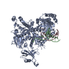
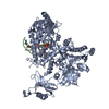
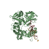
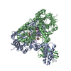
| Entry | Database: PDB / ID: 1m1l | ||||||
|---|---|---|---|---|---|---|---|
| Title | Human Suppressor of Fused (N-terminal domain) | ||||||
 Components Components | Suppressor of Fused | ||||||
 Keywords Keywords |  SIGNALING PROTEIN / Gene regulation / Hedgehog signaling / signal transduction / fused SIGNALING PROTEIN / Gene regulation / Hedgehog signaling / signal transduction / fused | ||||||
| Function / homology |  Function and homology information Function and homology informationsmoothened signaling pathway involved in ventral spinal cord interneuron specification / smoothened signaling pathway involved in spinal cord motor neuron cell fate specification / positive regulation of cellular response to drug / GLI-SUFU complex / ciliary tip / coronary vasculature development / aorta development / ventricular septum development / skin development / ciliary base ...smoothened signaling pathway involved in ventral spinal cord interneuron specification / smoothened signaling pathway involved in spinal cord motor neuron cell fate specification / positive regulation of cellular response to drug / GLI-SUFU complex / ciliary tip / coronary vasculature development / aorta development / ventricular septum development / skin development / ciliary base / negative regulation of protein import into nucleus / heart looping / spermatid development / negative regulation of osteoblast differentiation / Hedgehog 'off' state / negative regulation of smoothened signaling pathway / negative regulation of ubiquitin-dependent protein catabolic process / neural tube closure / Degradation of GLI1 by the proteasome / Hedgehog 'on' state / Degradation of GLI2 by the proteasome / GLI3 is processed to GLI3R by the proteasome / negative regulation of DNA-binding transcription factor activity / beta-catenin binding / transcription corepressor activity / regulation of DNA-templated transcription / protein kinase binding / negative regulation of transcription by RNA polymerase II / signal transduction / nucleoplasm / nucleus / cytosol / cytoplasmSimilarity search - Function | ||||||
| Biological species |  Homo sapiens (human) Homo sapiens (human) | ||||||
| Method | X-RAY DIFFRACTION / SYNCHROTRON / SAD / Resolution: 2.65 Å | ||||||
 Authors Authors | Merchant, M. / Vajdos, F.F. / Ultsch, M. / Maun, H.R. / Wendt, U. / Cannon, J. / Lazarus, R.A. / de Vos, A.M. / de Sauvage, F.J. | ||||||
 Citation Citation | Journal: Mol.Cell.Biol. / Year: 2004 Title: Suppressor of fused regulates Gli activity through a dual binding mechanism Authors: Merchant, M. / Vajdos, F.F. / Ultsch, M. / Maun, H.R. / Wendt, U. / Cannon, J. / Desmarais, W. / Lazarus, R.A. / de Vos, A.M. / de Sauvage, F.J. | ||||||
| History |
| ||||||
| Remark 300 | BIOMOLECULE: 1 THIS ENTRY CONTAINS THE CRYSTALLOGRAPHIC ASYMMETRIC UNIT REMARK 300 WHICH CONSISTS ... BIOMOLECULE: 1 THIS ENTRY CONTAINS THE CRYSTALLOGRAPHIC ASYMMETRIC UNIT REMARK 300 WHICH CONSISTS OF 4 CHAIN(S). AUTHOR STATES THAT BIOLOGICAL UNIT IS UNKNOWN. |
- Structure visualization
| Structure viewer | Molecule: MolmilJmol/JSmol |
|---|
- Downloads & links
-Download
| PDBx/mmCIF format | 1m1l.cif.gz | 197.1 KB | Display | PDBx/mmCIF format |
|---|---|---|---|---|
| PDB format | pdb1m1l.ent.gz | 160.7 KB | Display | PDB format |
| PDBx/mmJSON format | 1m1l.json.gz | Tree view | PDBx/mmJSON format | |
| Others |  Other downloads Other downloads |
-Validation report
| Arichive directory | https://data.pdbj.org/pub/pdb/validation_reports/m1/1m1lftp://data.pdbj.org/pub/pdb/validation_reports/m1/1m1l | HTTPS FTP |
|---|
-Related structure data
| Similar structure data |
|---|


-Links
 PDBj
PDBj

- Assembly
| Deposited unit | 
| ||||||||
|---|---|---|---|---|---|---|---|---|---|
| 1 |
| ||||||||
| Unit cell |
|
-Components
| #1: Protein | Mass: 26592.730 Da / Num. of mol.: 4 / Fragment: N-terminal domain (Residues 27-262) Source method: isolated from a genetically manipulated source Source: (gene. exp.) Homo sapiens (human) / Plasmid: pST-239 / Production host:  Escherichia coli (E. coli) / References: UniProt: Q9UMX1 Escherichia coli (E. coli) / References: UniProt: Q9UMX1#2: Water | ChemComp-HOH / | Water Mass: 18.015 Da / Num. of mol.: 346 / Source method: isolated from a natural source / Formula: H2O Mass: 18.015 Da / Num. of mol.: 346 / Source method: isolated from a natural source / Formula: H2O |
|---|
-Experimental details
-Experiment
| Experiment | Method: X-RAY DIFFRACTION / Number of used crystals: 1 |
|---|
- Sample preparation
| Crystal | Density Matthews: 3.94 Å3/Da / Density % sol: 68.77 % | |||||||||||||||||||||||||||||||||||||||||||||||||
|---|---|---|---|---|---|---|---|---|---|---|---|---|---|---|---|---|---|---|---|---|---|---|---|---|---|---|---|---|---|---|---|---|---|---|---|---|---|---|---|---|---|---|---|---|---|---|---|---|---|---|
| Crystal grow | Temperature: 279 K / Method: vapor diffusion, hanging drop / pH: 6 Details: 1 M LiCl, 0.1 M NaCitrate pH 5.0- 6.0, 10% w/v PEG 6000, VAPOR DIFFUSION, HANGING DROP, temperature 279K | |||||||||||||||||||||||||||||||||||||||||||||||||
| Crystal grow | *PLUS pH: 7.5 / Method: vapor diffusion, hanging drop | |||||||||||||||||||||||||||||||||||||||||||||||||
| Components of the solutions | *PLUS
|
-Data collection
| Diffraction | Mean temperature: 100 K |
|---|---|
| Diffraction source | Source: SYNCHROTRON / Site: SSRL  / Beamline: BL9-2 / Wavelength: 1 Å / Beamline: BL9-2 / Wavelength: 1 Å |
| Detector | Type: ADSC QUANTUM 4 / Detector: CCD / Date: May 15, 2001 |
| Radiation | Protocol: SINGLE WAVELENGTH / Monochromatic (M) / Laue (L): M / Scattering type: x-ray |
| Radiation wavelength | Wavelength: 1 Å / Relative weight: 1 |
| Reflection | Resolution: 2.65→29.81 Å / Num. all: 48643 / Num. obs: 48643 / % possible obs: 99.2 % / Observed criterion σ(I): -3 |
| Reflection | *PLUS Lowest resolution: 99 Å / Redundancy: 9.1 % / Num. measured all: 441505 / Rmerge(I) obs: 0.094 |
| Reflection shell | *PLUS Highest resolution: 2.65 Å / Lowest resolution: 2.7 Å / % possible obs: 100 % / Rmerge(I) obs: 0.362 |
- Processing
| Software |
| ||||||||||||||||
|---|---|---|---|---|---|---|---|---|---|---|---|---|---|---|---|---|---|
| Refinement | Method to determine structure: SAD / Resolution: 2.65→29.81 Å / Cross valid method: THROUGHOUT / σ(F): 0 / Stereochemistry target values: Engh & Huber
| ||||||||||||||||
| Refinement step | Cycle: LAST / Resolution: 2.65→29.81 Å
| ||||||||||||||||
| Refinement | *PLUS % reflection Rfree: 8 % / Rfactor Rfree: 0.256 / Rfactor Rwork: 0.224 | ||||||||||||||||
| Solvent computation | *PLUS | ||||||||||||||||
| Displacement parameters | *PLUS | ||||||||||||||||
| Refine LS restraints | *PLUS
|
