 Movie
Movie Controller

[English] 日本語
 Yorodumi
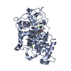
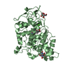
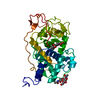
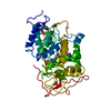
- PDB-1lyk: THE IMPACT OF THE PHYSICAL AND CHEMICAL ENVIROMENT ON THE MOLECUL... -
+ Open data

- Basic information
| Entry | Database: PDB / ID: 1lyk | ||||||
|---|---|---|---|---|---|---|---|
| Title | THE IMPACT OF THE PHYSICAL AND CHEMICAL ENVIROMENT ON THE MOLECULAR STRUCTURE OF COPRINUS CINEREUS PEROXIDASE | ||||||
 Components Components | Peroxidase | ||||||
 Keywords Keywords | OXIDOREDUCTASE / peroxidase / mutant / thermostability / Coprinus cinereus | ||||||
| Function / homology |  Function and homology informationlactoperoxidase activity / peroxidase / hydrogen peroxide catabolic process / cellular response to oxidative stress / heme binding / extracellular region / metal ion binding Function and homology informationlactoperoxidase activity / peroxidase / hydrogen peroxide catabolic process / cellular response to oxidative stress / heme binding / extracellular region / metal ion bindingSimilarity search - Function | ||||||
| Biological species |  Coprinopsis cinerea (fungus) Coprinopsis cinerea (fungus) | ||||||
| Method | X-RAY DIFFRACTION / MOLECULAR REPLACEMENT / Resolution: 2 Å | ||||||
 Authors Authors | Houborg, K. / Harris, P. / Petersen, J.F.W. / Rowland, P. / Poulsen, J.-C.N. / Schneider, P. / Vind, J. / Larsen, S. | ||||||
 Citation Citation | Journal: Acta Crystallogr.,Sect.D / Year: 2003 Title: Impact of the physical and chemical environment on the molecular structure of Coprinus cinereus peroxidase. Authors: Houborg, K. / Harris, P. / Petersen, J. / Rowland, P. / Poulsen, J.C. / Schneider, P. / Vind, J. / Larsen, S. | ||||||
| History |
|
- Structure visualization
| Structure viewer | Molecule: MolmilJmol/JSmol |
|---|
- Downloads & links
-Download
| PDBx/mmCIF format | 1lyk.cif.gz | 144.5 KB | Display | PDBx/mmCIF format |
|---|---|---|---|---|
| PDB format | pdb1lyk.ent.gz | 115.7 KB | Display | PDB format |
| PDBx/mmJSON format | 1lyk.json.gz | Tree view | PDBx/mmJSON format | |
| Others |  Other downloads Other downloads |
-Validation report
| Arichive directory | https://data.pdbj.org/pub/pdb/validation_reports/ly/1lykftp://data.pdbj.org/pub/pdb/validation_reports/ly/1lyk | HTTPS FTP |
|---|
-Related structure data


-Links
 PDBj
PDBj


- Assembly
| Deposited unit | 
| ||||||||||
|---|---|---|---|---|---|---|---|---|---|---|---|
| 1 | 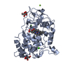
| ||||||||||
| 2 | 
| ||||||||||
| Unit cell |
| ||||||||||
| Components on special symmetry positions |
|
-Components
-Protein , 1 types, 2 molecules AB
| #1: Protein | / E.C.1.11.1.7 Mass: 35651.723 Da / Num. of mol.: 2 Source method: isolated from a genetically manipulated source Source: (gene. exp.) Coprinopsis cinerea (fungus) / Gene: CIP1 / Production host: Aspergillus oryzae (mold) / References: UniProt: P28314, peroxidase |
|---|
-Sugars , 3 types, 4 molecules 


| #2: Sugar | N-Acetylglucosamine Type: D-saccharide, beta linking / Mass: 221.208 Da / Num. of mol.: 2 Type: D-saccharide, beta linking / Mass: 221.208 Da / Num. of mol.: 2Source method: isolated from a genetically manipulated source Formula: C8H15NO6 #3: Sugar | ChemComp-BMA / | Mannose Type: D-saccharide, beta linking / Mass: 180.156 Da / Num. of mol.: 1 Type: D-saccharide, beta linking / Mass: 180.156 Da / Num. of mol.: 1Source method: isolated from a genetically manipulated source Formula: C6H12O6 #7: Sugar | ChemComp-MAN / | Mannose Type: D-saccharide, alpha linking / Mass: 180.156 Da / Num. of mol.: 1 Type: D-saccharide, alpha linking / Mass: 180.156 Da / Num. of mol.: 1Source method: isolated from a genetically manipulated source Formula: C6H12O6 |
|---|
-Non-polymers , 4 types, 483 molecules 


| #4: Chemical | ChemComp-CA /  Mass: 40.078 Da / Num. of mol.: 4 / Source method: obtained synthetically / Formula: Ca Mass: 40.078 Da / Num. of mol.: 4 / Source method: obtained synthetically / Formula: Ca#5: Chemical |  Mass: 24.305 Da / Num. of mol.: 2 / Source method: obtained synthetically / Formula: Mg Mass: 24.305 Da / Num. of mol.: 2 / Source method: obtained synthetically / Formula: Mg#6: Chemical | Heme B Mass: 616.487 Da / Num. of mol.: 2 / Source method: obtained synthetically / Formula: C34H32FeN4O4 Mass: 616.487 Da / Num. of mol.: 2 / Source method: obtained synthetically / Formula: C34H32FeN4O4#8: Water | ChemComp-HOH / | WaterMass: 18.015 Da / Num. of mol.: 475 / Source method: isolated from a natural source / Formula: H2O |
|---|
-Experimental details
-Experiment
| Experiment | Method: X-RAY DIFFRACTION / Number of used crystals: 1 |
|---|
- Sample preparation
| Crystal | Density Matthews: 2.63 Å3/Da / Density % sol: 53.31 % | ||||||||||||||||||||||||||||||
|---|---|---|---|---|---|---|---|---|---|---|---|---|---|---|---|---|---|---|---|---|---|---|---|---|---|---|---|---|---|---|---|
| Crystal grow | Temperature: 293 K / Method: vapor diffusion, hanging drop / pH: 7 Details: 18% PEG, 0.35M MgCl2, 10 mM KCN, pH 7.0, VAPOR DIFFUSION, HANGING DROP at 293K | ||||||||||||||||||||||||||||||
| Crystal grow | *PLUS Method: vapor diffusion | ||||||||||||||||||||||||||||||
| Components of the solutions | *PLUS
|
-Data collection
| Diffraction | Mean temperature: 288 K |
|---|---|
| Diffraction source | Source: ROTATING ANODE / Type: RIGAKU / Wavelength: 1.5418 Å |
| Detector | Type: RIGAKU RAXIS IIC / Detector: IMAGE PLATE / Date: Sep 20, 1994 |
| Radiation | Monochromator: graphite / Protocol: SINGLE WAVELENGTH / Monochromatic (M) / Laue (L): M / Scattering type: x-ray |
| Radiation wavelength | Wavelength: 1.5418 Å / Relative weight: 1 |
| Reflection | Resolution: 2→25 Å / Num. all: 49808 / Num. obs: 41462 / % possible obs: 83.3 % / Observed criterion σ(I): -3 / Rmerge(I) obs: 0.085 / Rsym value: 0.085 |
| Reflection shell | Resolution: 2→2.03 Å / Rmerge(I) obs: 0.49 / % possible all: 72.9 |
| Reflection | *PLUS Highest resolution: 2 Å / Lowest resolution: 25 Å / Num. measured all: 104109 |
| Reflection shell | *PLUS % possible obs: 72.9 % |
- Processing
| Software |
| ||||||||||||||||||||
|---|---|---|---|---|---|---|---|---|---|---|---|---|---|---|---|---|---|---|---|---|---|
| Refinement | Method to determine structure: MOLECULAR REPLACEMENT / Resolution: 2→25 Å / σ(F): 0 / σ(I): 0
| ||||||||||||||||||||
| Refinement step | Cycle: LAST / Resolution: 2→25 Å
| ||||||||||||||||||||
| Refine LS restraints |
| ||||||||||||||||||||
| LS refinement shell | Resolution: 2→2.09 Å /
| ||||||||||||||||||||
| Refinement | *PLUS Highest resolution: 2 Å / Lowest resolution: 25 Å / Rfactor obs: 0.179 | ||||||||||||||||||||
| Solvent computation | *PLUS | ||||||||||||||||||||
| Displacement parameters | *PLUS | ||||||||||||||||||||
| LS refinement shell | *PLUS Rfactor obs: 0.29 |
