[English] 日本語
Yorodumi
- PDB-1lyc: The impact of the physical and chemical enviroment on the molecul... -

+
Open data


ID or keywords:

Loading...

-
Basic information

Entry
Database: PDB / ID: 1lyc
TitleThe impact of the physical and chemical enviroment on the molecular structure of Coprinus cinereus peroxidase
ComponentsPeroxidase
KeywordsOXIDOREDUCTASE / peroxidase / mutant / thermostability / Coprinus cinereus
Function / homology
Function and homology information


lactoperoxidase activity / peroxidase / hydrogen peroxide catabolic process / cellular response to oxidative stress / heme binding / extracellular region / metal ion binding
Similarity search - Function
Fungal ligninase / Fungal ligninase, C-terminal / Fungal peroxidase extension region / Heme-binding peroxidase Ccp1-like / Peroxidase; domain 2 / Peroxidase, domain 2 / Peroxidase; domain 1 - #10 / Peroxidase; domain 1 / Peroxidases heam-ligand binding site / Peroxidase, active site ...Fungal ligninase / Fungal ligninase, C-terminal / Fungal peroxidase extension region / Heme-binding peroxidase Ccp1-like / Peroxidase; domain 2 / Peroxidase, domain 2 / Peroxidase; domain 1 - #10 / Peroxidase; domain 1 / Peroxidases heam-ligand binding site / Peroxidase, active site / Peroxidases active site signature. / Plant heme peroxidase family profile. / Haem peroxidase / Peroxidase / Peroxidases proximal heme-ligand signature. / Haem peroxidase superfamily / Orthogonal Bundle / Mainly Alpha
Similarity search - Domain/homology
PROTOPORPHYRIN IX CONTAINING FE / Peroxidase
Similarity search - Component
Biological speciesCoprinopsis cinerea (fungus)
MethodX-RAY DIFFRACTION / SYNCHROTRON / MOLECULAR REPLACEMENT / Resolution: 1.57 Å
AuthorsHouborg, K. / Harris, P. / Petersen, J.F.W. / Rowland, P. / Poulsen, J.-C.N. / Schneider, P. / Vind, J. / Larsen, S.
CitationJournal: Acta Crystallogr.,Sect.D / Year: 2003
Title: Impact of the physical and chemical environment on the molecular structure of Coprinus cinereus peroxidase.
Authors: Houborg, K. / Harris, P. / Petersen, J. / Rowland, P. / Poulsen, J.C. / Schneider, P. / Vind, J. / Larsen, S.
History
DepositionJun 7, 2002Deposition site: RCSB / Processing site: RCSB
Revision 1.0Jun 14, 2002Provider: repository / Type: Initial release
Revision 1.1Apr 28, 2008Group: Version format compliance
Revision 1.2Jul 13, 2011Group: Version format compliance
Revision 1.3Oct 27, 2021Group: Data collection / Database references / Derived calculations
Category: database_2 / diffrn_source ...database_2 / diffrn_source / pdbx_struct_conn_angle / struct_conn / struct_ref_seq_dif / struct_site
Item: _database_2.pdbx_DOI / _database_2.pdbx_database_accession ..._database_2.pdbx_DOI / _database_2.pdbx_database_accession / _diffrn_source.pdbx_synchrotron_site / _pdbx_struct_conn_angle.ptnr1_auth_comp_id / _pdbx_struct_conn_angle.ptnr1_auth_seq_id / _pdbx_struct_conn_angle.ptnr1_label_asym_id / _pdbx_struct_conn_angle.ptnr1_label_atom_id / _pdbx_struct_conn_angle.ptnr1_label_comp_id / _pdbx_struct_conn_angle.ptnr1_label_seq_id / _pdbx_struct_conn_angle.ptnr2_auth_comp_id / _pdbx_struct_conn_angle.ptnr2_auth_seq_id / _pdbx_struct_conn_angle.ptnr2_label_asym_id / _pdbx_struct_conn_angle.ptnr2_label_atom_id / _pdbx_struct_conn_angle.ptnr2_label_comp_id / _pdbx_struct_conn_angle.ptnr3_auth_comp_id / _pdbx_struct_conn_angle.ptnr3_auth_seq_id / _pdbx_struct_conn_angle.ptnr3_label_asym_id / _pdbx_struct_conn_angle.ptnr3_label_atom_id / _pdbx_struct_conn_angle.ptnr3_label_comp_id / _pdbx_struct_conn_angle.ptnr3_label_seq_id / _pdbx_struct_conn_angle.value / _struct_conn.pdbx_dist_value / _struct_conn.ptnr1_auth_comp_id / _struct_conn.ptnr1_auth_seq_id / _struct_conn.ptnr1_label_asym_id / _struct_conn.ptnr1_label_atom_id / _struct_conn.ptnr1_label_comp_id / _struct_conn.ptnr1_label_seq_id / _struct_conn.ptnr2_auth_comp_id / _struct_conn.ptnr2_auth_seq_id / _struct_conn.ptnr2_label_asym_id / _struct_conn.ptnr2_label_atom_id / _struct_conn.ptnr2_label_comp_id / _struct_conn.ptnr2_label_seq_id / _struct_ref_seq_dif.details / _struct_site.pdbx_auth_asym_id / _struct_site.pdbx_auth_comp_id / _struct_site.pdbx_auth_seq_id
Revision 1.4Apr 3, 2024Group: Data collection / Refinement description
Category: chem_comp_atom / chem_comp_bond / pdbx_initial_refinement_model

-
Structure visualization

Structure viewerMolecule:
MolmilJmol/JSmol

Downloads & links

-
Assembly

Deposited unit
A: Peroxidase
B: Peroxidase
hetero molecules


Theoretical massNumber of molelcules
Total (without water)72,5518
Polymers71,1572
Non-polymers1,3936
Water6,071337
1
A: Peroxidase
hetero molecules


Theoretical massNumber of molelcules
Total (without water)36,2754
Polymers35,5791
Non-polymers6973
Water181
TypeNameSymmetry operationNumber
identity operation1_555x,y,z1
2
B: Peroxidase
hetero molecules


Theoretical massNumber of molelcules
Total (without water)36,2754
Polymers35,5791
Non-polymers6973
Water181
TypeNameSymmetry operationNumber
identity operation1_555x,y,z1
Unit cell
Length a, b, c (Å)77.289, 115.055, 36.970
Angle α, β, γ (deg.)90.00, 90.00, 90.00
Int Tables number4
Cell settingmonoclinic
Space group name H-MP1211

-
Components

#1: Protein Peroxidase / / E.C.1.11.1.7


Mass: 35578.676 Da / Num. of mol.: 2
Source method: isolated from a genetically manipulated source
Source: (gene. exp.) Coprinopsis cinerea (fungus) / Gene: CIP1 / Production host: Aspergillus oryzae (mold) / References: UniProt: P28314, peroxidase
#2: Chemical
ChemComp-CA / CALCIUM ION


Mass: 40.078 Da / Num. of mol.: 4 / Source method: obtained synthetically / Formula: Ca
#3: Chemical ChemComp-HEM / PROTOPORPHYRIN IX CONTAINING FE / HEME / Heme B


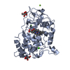
Mass: 616.487 Da / Num. of mol.: 2 / Source method: obtained synthetically / Formula: C34H32FeN4O4
#4: Water ChemComp-HOH / water / Water


Mass: 18.015 Da / Num. of mol.: 337 / Source method: isolated from a natural source / Formula: H2O

-
Experimental details

-
Experiment

ExperimentMethod: X-RAY DIFFRACTION / Number of used crystals: 3

-
Sample preparation

CrystalDensity Matthews: 2.31 Å3/Da / Density % sol: 46.71 %
Crystal growTemperature: 293 K / Method: vapor diffusion, hanging drop / pH: 4.25
Details: 1.5M-1.6M NH4H2PO, pH 4.0-4.5, VAPOR DIFFUSION, HANGING DROP, pH 4.25, temperature 293K
Crystal grow
*PLUS
Method: vapor diffusion / PH range low: 4.5 / PH range high: 4
Components of the solutions
*PLUS
IDConc.Common nameCrystal-IDSol-IDChemical formula
11.5-1.6 M1reservoirNH4H2PO4
217-21 mg/mlprotein1drop

-
Data collection

Diffraction
IDMean temperature (K)Crystal-ID
11001
21001
31001
Diffraction source
SourceSiteBeamlineTypeIDWavelength (Å)
SYNCHROTRONEMBL/DESY, HAMBURG BW7B10.8346
ROTATING ANODERIGAKU21.5418
ROTATING ANODERIGAKU31.5418
Detector
TypeIDDetectorDate
MARRESEARCH1IMAGE PLATESep 23, 1998
RIGAKU RAXIS IIC2IMAGE PLATEAug 27, 1999
MARRESEARCH3IMAGE PLATEFeb 25, 2000
Radiation
IDMonochromatorProtocolMonochromatic (M) / Laue (L)Scattering typeWavelength-ID
1mirrorsSINGLE WAVELENGTHMx-ray1
2graphiteSINGLE WAVELENGTHMx-ray1
3osmic mirrorsSINGLE WAVELENGTHMx-ray1
Radiation wavelength
IDWavelength (Å)Relative weight
10.83461
21.54181
ReflectionResolution: 1.57→15 Å / Num. all: 47148 / Num. obs: 46580 / % possible obs: 99 % / Observed criterion σ(I): -3 / Biso Wilson estimate: 28.9 Å2 / Rmerge(I) obs: 0.139 / Rsym value: 0.139
Reflection shellResolution: 1.57→1.6 Å / Rmerge(I) obs: 0.223 / Rsym value: 0.223 / % possible all: 96
Reflection
*PLUS
Highest resolution: 1.57 Å / Lowest resolution: 15 Å / Num. obs: 52771 / % possible obs: 88.2 % / Num. measured all: 155261 / Rmerge(I) obs: 0.073

-
Processing

Software
NameVersionClassification
DENZOdata reduction
SCALEPACKdata scaling
AMoREphasing
CNS0.9refinement
RefinementMethod to determine structure: MOLECULAR REPLACEMENT
Starting model: future PDB deposition

Resolution: 1.57→14.89 Å / Rfactor Rfree error: 0.005 / Isotropic thermal model: RESTRAINED / Cross valid method: THROUGHOUT / σ(F): 0 / σ(I): 0 / Stereochemistry target values: Engh & Huber
Details: The crystals are pseudo-merohedrally twinned. Therefore, they have been processsed in orthorhombic symmetry and the full monoclinic set has been generated hereafter, details will be described in the publication
RfactorNum. reflection% reflectionSelection details
Rfree0.265 4227 9.1 %RANDOM
Rwork0.223 ---
all0.225 89176 --
obs0.223 46580 98.3 %-
Solvent computationSolvent model: FLAT MODEL / Bsol: 60.5781 Å2 / ksol: 0.403111 e/Å3
Displacement parametersBiso mean: 34 Å2
Baniso -1Baniso -2Baniso -3
1--10.25 Å20 Å20 Å2
2---5.58 Å20 Å2
3---15.83 Å2
Refine analyzeLuzzati coordinate error free: 0.42 Å / Luzzati sigma a free: 0.63 Å
Refinement stepCycle: LAST / Resolution: 1.57→14.89 Å
ProteinNucleic acidLigandSolventTotal
Num. atoms4918 0 90 342 5350
Refine LS restraints
Refine-IDTypeDev idealDev ideal target
X-RAY DIFFRACTIONc_bond_d0.019
X-RAY DIFFRACTIONc_bond_d_na
X-RAY DIFFRACTIONc_bond_d_prot
X-RAY DIFFRACTIONc_angle_d
X-RAY DIFFRACTIONc_angle_d_na
X-RAY DIFFRACTIONc_angle_d_prot
X-RAY DIFFRACTIONc_angle_deg1.5
X-RAY DIFFRACTIONc_angle_deg_na
X-RAY DIFFRACTIONc_angle_deg_prot
X-RAY DIFFRACTIONc_dihedral_angle_d22
X-RAY DIFFRACTIONc_dihedral_angle_d_na
X-RAY DIFFRACTIONc_dihedral_angle_d_prot
X-RAY DIFFRACTIONc_improper_angle_d1
X-RAY DIFFRACTIONc_improper_angle_d_na
X-RAY DIFFRACTIONc_improper_angle_d_prot
X-RAY DIFFRACTIONc_mcbond_it0.781.5
X-RAY DIFFRACTIONc_mcangle_it1.252
X-RAY DIFFRACTIONc_scbond_it0.422
X-RAY DIFFRACTIONc_scangle_it0.672.5
LS refinement shellResolution: 1.57→1.67 Å / Rfactor Rfree error: 0.024 / Total num. of bins used: 6
RfactorNum. reflection% reflection
Rfree0.598 644 4.5 %
Rwork0.588 13763 -
obs--96.3 %
Xplor file
Refine-IDSerial noParam fileTopol file
X-RAY DIFFRACTION1PROTEIN_REP.PARAMPROTEIN.TOP
X-RAY DIFFRACTION2ION.PARAMION.TOP
X-RAY DIFFRACTION3WATER_REP.PARAMWATER.TOP
X-RAY DIFFRACTION4HEM_D.PARHEM_D.TOP
Refinement
*PLUS
Lowest resolution: 14.89 Å / Num. reflection obs: 86540
Solvent computation
*PLUS
Displacement parameters
*PLUS
Refine LS restraints
*PLUS
Refine-IDTypeDev ideal
X-RAY DIFFRACTIONc_dihedral_angle_d
X-RAY DIFFRACTIONc_dihedral_angle_deg22
X-RAY DIFFRACTIONc_improper_angle_d
X-RAY DIFFRACTIONc_improper_angle_deg1
LS refinement shell
*PLUS
Rfactor obs: 0.588

+
About Yorodumi

-
News

-
Feb 9, 2022. New format data for meta-information of EMDB entries

New format data for meta-information of EMDB entries

  • Version 3 of the EMDB header file is now the official format.
  • The previous official version 1.9 will be removed from the archive.

Related info.:EMDB header

External links:wwPDB to switch to version 3 of the EMDB data model

-
Aug 12, 2020. Covid-19 info

Covid-19 info

URL: https://pdbj.org/emnavi/covid19.php

New page: Covid-19 featured information page in EM Navigator.

Related info.:Covid-19 info / Mar 5, 2020. Novel coronavirus structure data

+
Mar 5, 2020. Novel coronavirus structure data

Novel coronavirus structure data

Related info.:Yorodumi Speices / Aug 12, 2020. Covid-19 info

External links:COVID-19 featured content - PDBj / Molecule of the Month (242):Coronavirus Proteases

+
Jan 31, 2019. EMDB accession codes are about to change! (news from PDBe EMDB page)

EMDB accession codes are about to change! (news from PDBe EMDB page)

  • The allocation of 4 digits for EMDB accession codes will soon come to an end. Whilst these codes will remain in use, new EMDB accession codes will include an additional digit and will expand incrementally as the available range of codes is exhausted. The current 4-digit format prefixed with “EMD-” (i.e. EMD-XXXX) will advance to a 5-digit format (i.e. EMD-XXXXX), and so on. It is currently estimated that the 4-digit codes will be depleted around Spring 2019, at which point the 5-digit format will come into force.
  • The EM Navigator/Yorodumi systems omit the EMD- prefix.

Related info.:Q: What is EMD? / ID/Accession-code notation in Yorodumi/EM Navigator

External links:EMDB Accession Codes are Changing Soon! / Contact to PDBj

+
Jul 12, 2017. Major update of PDB

Major update of PDB

  • wwPDB released updated PDB data conforming to the new PDBx/mmCIF dictionary.
  • This is a major update changing the version number from 4 to 5, and with Remediation, in which all the entries are updated.
  • In this update, many items about electron microscopy experimental information are reorganized (e.g. em_software).
  • Now, EM Navigator and Yorodumi are based on the updated data.

External links:wwPDB Remediation / Enriched Model Files Conforming to OneDep Data Standards Now Available in the PDB FTP Archive

-
Yorodumi

Thousand views of thousand structures

  • Yorodumi is a browser for structure data from EMDB, PDB, SASBDB, etc.
  • This page is also the successor to EM Navigator detail page, and also detail information page/front-end page for Omokage search.
  • The word "yorodu" (or yorozu) is an old Japanese word meaning "ten thousand". "mi" (miru) is to see.

Related info.:EMDB / PDB / SASBDB / Comparison of 3 databanks / Yorodumi Search / Aug 31, 2016. New EM Navigator & Yorodumi / Yorodumi Papers / Jmol/JSmol / Function and homology information / Changes in new EM Navigator and Yorodumi

Read more