 Movie
Movie Controller

[English] 日本語
 Yorodumi
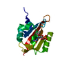
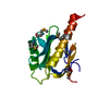
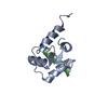
- PDB-1lxf: Structure of the Regulatory N-domain of Human Cardiac Troponin C ... -
+ Open data

- Basic information
| Entry | Database: PDB / ID: 1lxf | ||||||
|---|---|---|---|---|---|---|---|
| Title | Structure of the Regulatory N-domain of Human Cardiac Troponin C in Complex with Human Cardiac Troponin-I(147-163) and Bepridil | ||||||
 Components Components |
| ||||||
 Keywords Keywords | METAL BINDING PROTEIN /  PROTEIN BINDING / muscle / cardiac troponin C-drug interaction / bepridil / cardiac troponin I-drug interaction PROTEIN BINDING / muscle / cardiac troponin C-drug interaction / bepridil / cardiac troponin I-drug interaction | ||||||
| Function / homology |  Function and homology information Function and homology informationregulation of systemic arterial blood pressure by ischemic conditions / regulation of muscle filament sliding speed / troponin T binding / diaphragm contraction / troponin C binding / regulation of ATP-dependent activity / cardiac Troponin complex / cardiac myofibril / regulation of smooth muscle contraction / troponin complex ...regulation of systemic arterial blood pressure by ischemic conditions / regulation of muscle filament sliding speed / troponin T binding / diaphragm contraction / troponin C binding / regulation of ATP-dependent activity / cardiac Troponin complex / cardiac myofibril / regulation of smooth muscle contraction / troponin complex / regulation of muscle contraction / transition between fast and slow fiber / negative regulation of ATP-dependent activity / Striated Muscle Contraction / response to metal ion / regulation of cardiac muscle contraction by calcium ion signaling / myosin II complex / ventricular cardiac muscle tissue morphogenesis / heart contraction / troponin I binding / skeletal muscle contraction / calcium channel inhibitor activity / vasculogenesis / cardiac muscle contraction / Ion homeostasis / sarcomere / intracellular calcium ion homeostasis / calcium-dependent protein binding / actin filament binding / actin binding / heart development / protein domain specific binding / calcium ion binding / protein kinase binding / protein homodimerization activity / cytosolSimilarity search - Function | ||||||
| Biological species |  Homo sapiens (human) Homo sapiens (human) | ||||||
| Method | SOLUTION NMR / simulated annealing | ||||||
 Authors Authors | Wang, X. / Li, M.X. / Sykes, B.D. | ||||||
 Citation Citation | Journal: J.Biol.Chem. / Year: 2002 Title: Structure of the regulatory N-domain of human cardiac troponin C in complex with human cardiac troponin I147-163 and bepridil. Authors: Wang, X. / Li, M.X. / Sykes, B.D. | ||||||
| History |
|
- Structure visualization
| Structure viewer | Molecule: MolmilJmol/JSmol |
|---|
- Downloads & links
-Download
| PDBx/mmCIF format | 1lxf.cif.gz | 981.1 KB | Display | PDBx/mmCIF format |
|---|---|---|---|---|
| PDB format | pdb1lxf.ent.gz | 829.4 KB | Display | PDB format |
| PDBx/mmJSON format | 1lxf.json.gz | Tree view | PDBx/mmJSON format | |
| Others |  Other downloads Other downloads |
-Validation report
| Arichive directory | https://data.pdbj.org/pub/pdb/validation_reports/lx/1lxfftp://data.pdbj.org/pub/pdb/validation_reports/lx/1lxf | HTTPS FTP |
|---|
-Related structure data
| Similar structure data |
|---|


-Links
 PDBj
PDBj


- Assembly
| Deposited unit | 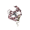
| |||||||||
|---|---|---|---|---|---|---|---|---|---|---|
| 1 |
| |||||||||
| NMR ensembles |
|
-Components
| #1: Protein | / Cardiac Troponin C / TN-C Mass: 10070.304 Da / Num. of mol.: 1 / Fragment: Regulatory N Domain (residues 1-89) Source method: isolated from a genetically manipulated source Source: (gene. exp.) Homo sapiens (human) / Gene: TNNC / Plasmid: pET-3a / Production host:  Escherichia coli (E. coli) / Strain (production host): BL21-(DE3plysS) / References: UniProt: P63316 Escherichia coli (E. coli) / Strain (production host): BL21-(DE3plysS) / References: UniProt: P63316 |
|---|---|
| #2: Protein/peptide | Mass: 1806.183 Da / Num. of mol.: 1 / Fragment: Switch Peptide (residues 147-163) / Source method: obtained synthetically Details: The sequence of the protein is naturally found in Homo sapiens. The protein was chemically synthesized. References: UniProt: P19429 |
| #3: Chemical | ChemComp-CA /   Mass: 40.078 Da / Num. of mol.: 1 / Source method: obtained synthetically / Formula: Ca Mass: 40.078 Da / Num. of mol.: 1 / Source method: obtained synthetically / Formula: Ca |
| #4: Chemical | ChemComp-BEP / Bepridil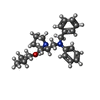  Mass: 366.540 Da / Num. of mol.: 1 / Source method: obtained synthetically / Formula: C24H34N2O / Comment: channel blocker*YM Mass: 366.540 Da / Num. of mol.: 1 / Source method: obtained synthetically / Formula: C24H34N2O / Comment: channel blocker*YM |
-Experimental details
-Experiment
| Experiment | Method: SOLUTION NMR | ||||||||||||||||||||||||||||||||||||||||
|---|---|---|---|---|---|---|---|---|---|---|---|---|---|---|---|---|---|---|---|---|---|---|---|---|---|---|---|---|---|---|---|---|---|---|---|---|---|---|---|---|---|
| NMR experiment |
|
- Sample preparation
| Details | Contents: 1mM cNTnC, 3mM cTnI147-163, 1.5mM bepridil, 5mM DTT, 100mM KCl, 10mM IMDZ, trace amount of NaN3, 50uM DSS, 90% H2O, 10% D2O Solvent system: 90% H2O/10% D2O |
|---|---|
| Sample conditions | Ionic strength: 0.1 / pH: 6.7 / Pressure: ambient / Temperature: 303 K |
| Crystal grow | *PLUS Method: other / Details: NMR |
-NMR measurement
| Radiation | Protocol: SINGLE WAVELENGTH / Monochromatic (M) / Laue (L): M | ||||||||||||||||||||
|---|---|---|---|---|---|---|---|---|---|---|---|---|---|---|---|---|---|---|---|---|---|
| Radiation wavelength | Relative weight: 1 | ||||||||||||||||||||
| NMR spectrometer |
|
- Processing
| NMR software |
| ||||||||||||||||
|---|---|---|---|---|---|---|---|---|---|---|---|---|---|---|---|---|---|
| Refinement | Method: simulated annealing / Software ordinal: 1 Details: A total of 1169 NOE restraints, 88 dihedral angle restraints used for the refinement of cNTnC, 30 NOE restraints used between cNTnC and cTnI147-163, 28 NOE restraints used between cNTnC and ...Details: A total of 1169 NOE restraints, 88 dihedral angle restraints used for the refinement of cNTnC, 30 NOE restraints used between cNTnC and cTnI147-163, 28 NOE restraints used between cNTnC and bepridil, 24 intramolecular NOE restraints and 12 dihedral angle restraints used within cTnI147-163. | ||||||||||||||||
| NMR representative | Selection criteria: lowest energy | ||||||||||||||||
| NMR ensemble | Conformer selection criteria: all calculated structures submitted Conformers calculated total number: 30 / Conformers submitted total number: 30 |
