 Movie
Movie Controller


+ Open data

- Basic information
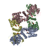
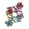
| Entry | Database: PDB / ID: 1ltq | ||||||
|---|---|---|---|---|---|---|---|
| Title | CRYSTAL STRUCTURE OF T4 POLYNUCLEOTIDE KINASE | ||||||
 Components Components | POLYNUCLEOTIDE KINASE Polynucleotide 5'-hydroxyl-kinase Polynucleotide 5'-hydroxyl-kinase | ||||||
 Keywords Keywords | TRANSFERASE / KINASE / PHOSPHATASE / ALPHA/BETA / P-LOOP | ||||||
| Function / homology |  Function and homology informationdeoxynucleotide 3'-phosphatase / deoxynucleotide 3'-phosphatase activity / polynucleotide 5'-hydroxyl-kinase / ATP-dependent polydeoxyribonucleotide 5'-hydroxyl-kinase activity / phosphorylation / DNA repair / ATP binding Function and homology informationdeoxynucleotide 3'-phosphatase / deoxynucleotide 3'-phosphatase activity / polynucleotide 5'-hydroxyl-kinase / ATP-dependent polydeoxyribonucleotide 5'-hydroxyl-kinase activity / phosphorylation / DNA repair / ATP bindingSimilarity search - Function | ||||||
| Biological species |  Enterobacteria phage T4 (virus) Enterobacteria phage T4 (virus) | ||||||
| Method | X-RAY DIFFRACTION / SYNCHROTRON / SAD / Resolution: 2.33 Å | ||||||
 Authors Authors | Galburt, E.A. / Pelletier, J. / Wilson, G. / Stoddard, B.L. | ||||||
 Citation Citation | Journal: Structure / Year: 2002 Title: Structure of a tRNA repair enzyme and molecular biology workhorse: T4 polynucleotide kinase. Authors: Galburt, E.A. / Pelletier, J. / Wilson, G. / Stoddard, B.L. | ||||||
| History |
|
- Structure visualization
| Structure viewer | Molecule: MolmilJmol/JSmol |
|---|
- Downloads & links
-Download
| PDBx/mmCIF format | 1ltq.cif.gz | 72 KB | Display | PDBx/mmCIF format |
|---|---|---|---|---|
| PDB format | pdb1ltq.ent.gz | 57.8 KB | Display | PDB format |
| PDBx/mmJSON format | 1ltq.json.gz | Tree view | PDBx/mmJSON format | |
| Others |  Other downloads Other downloads |
-Validation report
| Arichive directory | https://data.pdbj.org/pub/pdb/validation_reports/lt/1ltqftp://data.pdbj.org/pub/pdb/validation_reports/lt/1ltq | HTTPS FTP |
|---|
-Related structure data
| Similar structure data |
|---|

-Links
 PDBj
PDBj- Assembly
| Deposited unit | 
| ||||||||
|---|---|---|---|---|---|---|---|---|---|
| 1 | 
| ||||||||
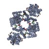
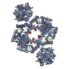
| Unit cell |
|
-Components
| #1: Protein | Polynucleotide 5'-hydroxyl-kinase / PNK / Polynucleotide 5'-hydroxyl-kinase Mass: 35234.594 Da / Num. of mol.: 1 Source method: isolated from a genetically manipulated source Source: (gene. exp.) Enterobacteria phage T4 (virus) / Genus: T4-like viruses / Species: Enterobacteria phage T4 sensu lato / Production host:  Escherichia coli (E. coli) Escherichia coli (E. coli)References: UniProt: P06855, polynucleotide 5'-hydroxyl-kinase |
|---|---|
| #2: Chemical | ChemComp-ADP / Adenosine diphosphate  Mass: 427.201 Da / Num. of mol.: 1 / Source method: obtained synthetically / Formula: C10H15N5O10P2 / Comment: ADP, energy-carrying molecule*YM Mass: 427.201 Da / Num. of mol.: 1 / Source method: obtained synthetically / Formula: C10H15N5O10P2 / Comment: ADP, energy-carrying molecule*YM |
| #3: Chemical | ChemComp-DMS / Dimethyl sulfoxide  Mass: 78.133 Da / Num. of mol.: 1 / Source method: obtained synthetically / Formula: C2H6OS / Comment: DMSO, precipitant*YM Mass: 78.133 Da / Num. of mol.: 1 / Source method: obtained synthetically / Formula: C2H6OS / Comment: DMSO, precipitant*YM |
| #4: Water | ChemComp-HOH / Water Mass: 18.015 Da / Num. of mol.: 144 / Source method: isolated from a natural source / Formula: H2O Mass: 18.015 Da / Num. of mol.: 144 / Source method: isolated from a natural source / Formula: H2O |
-Experimental details
-Experiment
| Experiment | Method: X-RAY DIFFRACTION / Number of used crystals: 1 |
|---|
- Sample preparation
| Crystal | Density Matthews: 3.24 Å3/Da / Density % sol: 62.06 % | |||||||||||||||||||||||||||||||||||||||||||||||||||||||||||||||
|---|---|---|---|---|---|---|---|---|---|---|---|---|---|---|---|---|---|---|---|---|---|---|---|---|---|---|---|---|---|---|---|---|---|---|---|---|---|---|---|---|---|---|---|---|---|---|---|---|---|---|---|---|---|---|---|---|---|---|---|---|---|---|---|---|
| Crystal grow | Temperature: 298 K / Method: vapor diffusion, hanging drop / pH: 6.5 Details: PEG 4000, potassium chloride, MES, Tris, ATP, DTT, EDTA, pH 6.5, VAPOR DIFFUSION, HANGING DROP, temperature 298K | |||||||||||||||||||||||||||||||||||||||||||||||||||||||||||||||
| Crystal grow | *PLUS pH: 7.4 | |||||||||||||||||||||||||||||||||||||||||||||||||||||||||||||||
| Components of the solutions | *PLUS
|
-Data collection
| Diffraction | Mean temperature: 100 K |
|---|---|
| Diffraction source | Source: SYNCHROTRON / Site: ALS  / Beamline: 5.0.2 / Wavelength: 0.97873 Å / Beamline: 5.0.2 / Wavelength: 0.97873 Å |
| Detector | Type: ADSC QUANTUM 4 / Detector: CCD / Date: Mar 10, 2002 |
| Radiation | Monochromator: Double-crystal Si(111) / Protocol: MAD / Monochromatic (M) / Laue (L): M / Scattering type: x-ray |
| Radiation wavelength | Wavelength: 0.97873 Å / Relative weight: 1 |
| Reflection | Resolution: 2.3→50 Å / Num. all: 38288 / Num. obs: 38288 / % possible obs: 99.1 % / Observed criterion σ(F): 0 / Observed criterion σ(I): 0 / Redundancy: 5 % / Biso Wilson estimate: 30 Å2 / Rmerge(I) obs: 0.073 / Net I/σ(I): 12.8 |
| Reflection shell | Resolution: 2.3→2.38 Å / Rmerge(I) obs: 0.288 / Mean I/σ(I) obs: 5.7 / % possible all: 97.7 |
| Reflection | *PLUS Highest resolution: 2.3 Å / Lowest resolution: 50 Å / Num. measured all: 191084 / Rmerge(I) obs: 0.073 |
| Reflection shell | *PLUS % possible obs: 97.7 % / Rmerge(I) obs: 0.288 |
- Processing
| Software |
| |||||||||||||||||||||||||
|---|---|---|---|---|---|---|---|---|---|---|---|---|---|---|---|---|---|---|---|---|---|---|---|---|---|---|
| Refinement | Method to determine structure: SAD / Resolution: 2.33→50 Å / Cross valid method: THROUGHOUT / σ(F): 0 / Stereochemistry target values: Engh & Huber
| |||||||||||||||||||||||||
| Refine analyze | Luzzati coordinate error obs: 0.3 Å | |||||||||||||||||||||||||
| Refinement step | Cycle: LAST / Resolution: 2.33→50 Å
| |||||||||||||||||||||||||
| Refinement | *PLUS Rfactor all: 0.238 / Rfactor Rfree: 0.261 / Rfactor Rwork: 0.236 | |||||||||||||||||||||||||
| Solvent computation | *PLUS | |||||||||||||||||||||||||
| Displacement parameters | *PLUS | |||||||||||||||||||||||||
| Refine LS restraints | *PLUS
|
