 Movie
Movie Controller

[English] 日本語
 Yorodumi
- PDB-1lm7: Structures of two intermediate filament-binding fragments of desm... -
+ Open data

- Basic information
| Entry | Database: PDB / ID: 1lm7 | ||||||
|---|---|---|---|---|---|---|---|
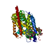
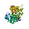
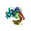
| Title | Structures of two intermediate filament-binding fragments of desmoplakin reveal a unique repeat motif structure | ||||||
 Components Components | subdomain of Desmoplakin Carboxy-Terminal domain (DPCT) | ||||||
 Keywords Keywords |  STRUCTURAL PROTEIN / plakin repeat STRUCTURAL PROTEIN / plakin repeat | ||||||
| Function / homology |  Function and homology information Function and homology informationbundle of His cell-Purkinje myocyte adhesion involved in cell communication / cell adhesive protein binding involved in bundle of His cell-Purkinje myocyte communication / desmosome organization / Keratinization / protein localization to cell-cell junction / ventricular compact myocardium morphogenesis / desmosome / intermediate filament organization / epithelial cell-cell adhesion / intermediate filament cytoskeleton organization ...bundle of His cell-Purkinje myocyte adhesion involved in cell communication / cell adhesive protein binding involved in bundle of His cell-Purkinje myocyte communication / desmosome organization / Keratinization / protein localization to cell-cell junction / ventricular compact myocardium morphogenesis / desmosome / intermediate filament organization / epithelial cell-cell adhesion / intermediate filament cytoskeleton organization / Formation of the cornified envelope / peptide cross-linking / fascia adherens / cornified envelope / regulation of ventricular cardiac muscle cell action potential / Apoptotic cleavage of cell adhesion proteins / adherens junction organization / RND1 GTPase cycle / RND3 GTPase cycle / skin development / intermediate filament / regulation of heart rate by cardiac conduction / ficolin-1-rich granule membrane / intercalated disc / epidermis development / keratinocyte differentiation / protein kinase C binding / adherens junction / wound healing / structural constituent of cytoskeleton / cell-cell adhesion / scaffold protein binding / basolateral plasma membrane / Neutrophil degranulation / structural molecule activity / RNA binding / extracellular exosome / nucleus / plasma membrane / cytoplasmSimilarity search - Function | ||||||
| Biological species |  Homo sapiens (human) Homo sapiens (human) | ||||||
| Method | X-RAY DIFFRACTION / SYNCHROTRON / SAD / Resolution: 3 Å | ||||||
 Authors Authors | Choi, H.J. / Park-Snyder, S. / Pascoe, L.T. / Green, K.J. / Weis, W.I. | ||||||
 Citation Citation | Journal: Nat.Struct.Biol. / Year: 2002 Title: Structures of two intermediate filament-binding fragments of desmoplakin reveal a unique repeat motif structure. Authors: Choi, H.J. / Park-Snyder, S. / Pascoe, L.T. / Green, K.J. / Weis, W.I. | ||||||
| History |
|
- Structure visualization
| Structure viewer | Molecule: MolmilJmol/JSmol |
|---|
- Downloads & links
-Download
| PDBx/mmCIF format | 1lm7.cif.gz | 100.7 KB | Display | PDBx/mmCIF format |
|---|---|---|---|---|
| PDB format | pdb1lm7.ent.gz | 78.7 KB | Display | PDB format |
| PDBx/mmJSON format | 1lm7.json.gz | Tree view | PDBx/mmJSON format | |
| Others |  Other downloads Other downloads |
-Validation report
| Arichive directory | https://data.pdbj.org/pub/pdb/validation_reports/lm/1lm7ftp://data.pdbj.org/pub/pdb/validation_reports/lm/1lm7 | HTTPS FTP |
|---|
-Related structure data


-Links
 PDBj
PDBj

- Assembly
| Deposited unit | 
| ||||||||
|---|---|---|---|---|---|---|---|---|---|
| 1 |
| ||||||||
| Unit cell |
|
-Components
| #1: Protein | Mass: 27569.258 Da / Num. of mol.: 2 / Fragment: residues 2209-2456 Source method: isolated from a genetically manipulated source Source: (gene. exp.) Homo sapiens (human) / Plasmid: pPROEX HTc / Production host:  Escherichia coli (E. coli) / Strain (production host): DH5a / References: UniProt: P15924 Escherichia coli (E. coli) / Strain (production host): DH5a / References: UniProt: P15924 |
|---|
-Experimental details
-Experiment
| Experiment | Method: X-RAY DIFFRACTION / Number of used crystals: 1 |
|---|
- Sample preparation
| Crystal | Density Matthews: 2.24 Å3/Da / Density % sol: 45.06 % | ||||||||||||||||||||||||
|---|---|---|---|---|---|---|---|---|---|---|---|---|---|---|---|---|---|---|---|---|---|---|---|---|---|
| Crystal grow | Temperature: 293 K / Method: vapor diffusion, hanging drop / pH: 6 Details: PEG1500, MES, pH 6.0, VAPOR DIFFUSION, HANGING DROP, temperature 293K | ||||||||||||||||||||||||
| Crystal grow | *PLUS Temperature: 20 ℃ | ||||||||||||||||||||||||
| Components of the solutions | *PLUS
|
-Data collection
| Diffraction | Mean temperature: 100 K |
|---|---|
| Diffraction source | Source: SYNCHROTRON / Site: SSRL  / Beamline: BL9-2 / Wavelength: 0.979 Å / Beamline: BL9-2 / Wavelength: 0.979 Å |
| Detector | Type: ADSC QUANTUM 4 / Detector: CCD / Date: May 7, 2001 / Details: mirrors |
| Radiation | Monochromator: graphite / Protocol: SINGLE WAVELENGTH / Monochromatic (M) / Laue (L): M / Scattering type: x-ray |
| Radiation wavelength | Wavelength: 0.979 Å / Relative weight: 1 |
| Reflection | Resolution: 2.8→50 Å / Num. all: 11017 / Num. obs: 10670 / % possible obs: 93.8 % / Observed criterion σ(F): 0 / Observed criterion σ(I): -3 / Rmerge(I) obs: 0.088 |
| Reflection shell | Resolution: 3→3.11 Å / Rmerge(I) obs: 0.232 / % possible all: 84.3 |
| Reflection | *PLUS Highest resolution: 3 Å / Lowest resolution: 50 Å / Num. obs: 9183 / Num. measured all: 208172 / Rmerge(I) obs: 0.087 |
| Reflection shell | *PLUS % possible obs: 84.3 % / Rmerge(I) obs: 0.23 / Mean I/σ(I) obs: 15.2 |
- Processing
| Software |
| ||||||||||||||||||||||||||||||||||||||||||||||||||||||||||||||||||||||||||||||||
|---|---|---|---|---|---|---|---|---|---|---|---|---|---|---|---|---|---|---|---|---|---|---|---|---|---|---|---|---|---|---|---|---|---|---|---|---|---|---|---|---|---|---|---|---|---|---|---|---|---|---|---|---|---|---|---|---|---|---|---|---|---|---|---|---|---|---|---|---|---|---|---|---|---|---|---|---|---|---|---|---|---|
| Refinement | Method to determine structure: SAD / Resolution: 3→35.74 Å / Rfactor Rfree error: 0.01 / Data cutoff high absF: 322205.39 / Data cutoff high rms absF: 322205.39 / Data cutoff low absF: 0 / Isotropic thermal model: RESTRAINED / Cross valid method: THROUGHOUT / σ(F): 0 / Stereochemistry target values: Engh & Huber
| ||||||||||||||||||||||||||||||||||||||||||||||||||||||||||||||||||||||||||||||||
| Solvent computation | Solvent model: FLAT MODEL / Bsol: 27.4838 Å2 / ksol: 0.301884 e/Å3 | ||||||||||||||||||||||||||||||||||||||||||||||||||||||||||||||||||||||||||||||||
| Displacement parameters | Biso mean: 40.5 Å2
| ||||||||||||||||||||||||||||||||||||||||||||||||||||||||||||||||||||||||||||||||
| Refine analyze |
| ||||||||||||||||||||||||||||||||||||||||||||||||||||||||||||||||||||||||||||||||
| Refinement step | Cycle: LAST / Resolution: 3→35.74 Å
| ||||||||||||||||||||||||||||||||||||||||||||||||||||||||||||||||||||||||||||||||
| Refine LS restraints |
| ||||||||||||||||||||||||||||||||||||||||||||||||||||||||||||||||||||||||||||||||
| Refine LS restraints NCS | NCS model details: CONSTR | ||||||||||||||||||||||||||||||||||||||||||||||||||||||||||||||||||||||||||||||||
| LS refinement shell | Resolution: 3→3.19 Å / Rfactor Rfree error: 0.033 / Total num. of bins used: 6
| ||||||||||||||||||||||||||||||||||||||||||||||||||||||||||||||||||||||||||||||||
| Xplor file | Serial no: 1 / Param file: PROTEIN_REP.PARAM / Topol file: PROTEIN.TOP | ||||||||||||||||||||||||||||||||||||||||||||||||||||||||||||||||||||||||||||||||
| Refinement | *PLUS Highest resolution: 3 Å / Lowest resolution: 100 Å / % reflection Rfree: 10 % | ||||||||||||||||||||||||||||||||||||||||||||||||||||||||||||||||||||||||||||||||
| Solvent computation | *PLUS | ||||||||||||||||||||||||||||||||||||||||||||||||||||||||||||||||||||||||||||||||
| Displacement parameters | *PLUS | ||||||||||||||||||||||||||||||||||||||||||||||||||||||||||||||||||||||||||||||||
| Refine LS restraints | *PLUS
|
