 Movie
Movie Controller

[English] 日本語
 Yorodumi
- PDB-1l6z: CRYSTAL STRUCTURE OF MURINE CEACAM1A[1,4]: A CORONAVIRUS RECEPTOR... -
+ Open data

- Basic information
| Entry | Database: PDB / ID: 1l6z | |||||||||
|---|---|---|---|---|---|---|---|---|---|---|
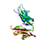
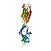
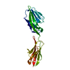
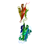
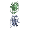
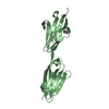
| Title | CRYSTAL STRUCTURE OF MURINE CEACAM1A[1,4]: A CORONAVIRUS RECEPTOR AND CELL ADHESION MOLECULE IN THE CEA FAMILY | |||||||||
 Components Components | biliary glycoprotein C | |||||||||
 Keywords Keywords |  CELL ADHESION / Ig-like domain / CEA Family / Coronavirus Receptor CELL ADHESION / Ig-like domain / CEA Family / Coronavirus Receptor | |||||||||
| Function / homology |  Function and homology information Function and homology informationFibronectin matrix formation / granulocyte colony-stimulating factor receptor binding / positive regulation of homophilic cell adhesion / Post-translational modification: synthesis of GPI-anchored proteins / GPI anchor binding / regulation of endothelial cell differentiation / insulin receptor internalization / negative regulation of cytotoxic T cell degranulation / granulocyte colony-stimulating factor signaling pathway / regulation of homophilic cell adhesion ...Fibronectin matrix formation / granulocyte colony-stimulating factor receptor binding / positive regulation of homophilic cell adhesion / Post-translational modification: synthesis of GPI-anchored proteins / GPI anchor binding / regulation of endothelial cell differentiation / insulin receptor internalization / negative regulation of cytotoxic T cell degranulation / granulocyte colony-stimulating factor signaling pathway / regulation of homophilic cell adhesion / regulation of epidermal growth factor receptor signaling pathway / regulation of sprouting angiogenesis / regulation of blood vessel remodeling / negative regulation of hepatocyte proliferation / negative regulation of natural killer cell mediated cytotoxicity directed against tumor cell target / transforming growth factor beta ligand-receptor complex / negative regulation of lipid biosynthetic process / bile acid transmembrane transporter activity / negative regulation of T cell mediated cytotoxicity / regulation of endothelial cell migration / filamin binding / negative regulation of granulocyte differentiation / insulin catabolic process / Cell surface interactions at the vascular wall / common myeloid progenitor cell proliferation / Toll-like receptor binding / negative regulation of interleukin-1 production / negative regulation of fatty acid biosynthetic process / positive regulation of vasculogenesis / cell-cell adhesion via plasma-membrane adhesion molecules / regulation of immune system process / cell-cell junction organization / negative regulation of platelet aggregation / bile acid and bile salt transport / negative regulation of vascular permeability / negative regulation of bone resorption / negative regulation of cytokine production / wound healing, spreading of cells / ciliary membrane / microvillus membrane / negative regulation of T cell receptor signaling pathway / regulation of phosphatidylinositol 3-kinase/protein kinase B signal transduction / blood vessel development / negative regulation of osteoclast differentiation / lateral plasma membrane / transport vesicle / negative regulation of T cell proliferation / protein tyrosine kinase binding / Neutrophil degranulation / regulation of ERK1 and ERK2 cascade / basal plasma membrane / regulation of cell growth / adherens junction / negative regulation of protein kinase activity / kinase binding / cellular response to insulin stimulus / cell-cell junction / virus receptor activity / cell junction / actin binding / basolateral plasma membrane / protein phosphatase binding / protein dimerization activity / calmodulin binding / cell adhesion / apical plasma membrane / protein heterodimerization activity / external side of plasma membrane / protein-containing complex binding / protein kinase binding / cell surface / signal transduction / protein homodimerization activity / extracellular space / membrane / identical protein binding / plasma membraneSimilarity search - Function | |||||||||
| Biological species |  Mus musculus (house mouse) Mus musculus (house mouse) | |||||||||
| Method | X-RAY DIFFRACTION / SYNCHROTRON / MAD, Molecular Replacement / Resolution: 3.32 Å | |||||||||
 Authors Authors | Tan, K. / Zelus, B.D. / Meijers, R. / Liu, J.-H. / Bergelson, J.M. / Duke, N. / Zhang, R. / Joachimiak, A. / Holmes, K.V. / Wang, J.-H. | |||||||||
 Citation Citation | Journal: Embo J. / Year: 2002 Title: CRYSTAL STRUCTURE OF MURINE sCEACAM1a[1,4]: A CORONAVIRUS RECEPTOR IN THE CEA FAMILY Authors: Tan, K. / Zelus, B.D. / Meijers, R. / Liu, J.-H. / Bergelson, J.M. / Duke, N. / Zhang, R. / Joachimiak, A. / Holmes, K.V. / Wang, J.-H. | |||||||||
| History |
|
- Structure visualization
| Structure viewer | Molecule: MolmilJmol/JSmol |
|---|
- Downloads & links
-Download
| PDBx/mmCIF format | 1l6z.cif.gz | 59.3 KB | Display | PDBx/mmCIF format |
|---|---|---|---|---|
| PDB format | pdb1l6z.ent.gz | 41.3 KB | Display | PDB format |
| PDBx/mmJSON format | 1l6z.json.gz | Tree view | PDBx/mmJSON format | |
| Others |  Other downloads Other downloads |
-Validation report
| Arichive directory | https://data.pdbj.org/pub/pdb/validation_reports/l6/1l6zftp://data.pdbj.org/pub/pdb/validation_reports/l6/1l6z | HTTPS FTP |
|---|
-Related structure data


-Links
 PDBj
PDBj


- Assembly
| Deposited unit | 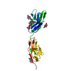
| ||||||||
|---|---|---|---|---|---|---|---|---|---|
| 1 |
| ||||||||
| Unit cell |
|
-Components
| #1: Protein | Mass: 24533.693 Da / Num. of mol.: 1 Source method: isolated from a genetically manipulated source Source: (gene. exp.) Mus musculus (house mouse) / Gene: Murine CEACAM1A / Cell (production host): OVARY CELLS / Production host:  Cricetulus griseus (Chinese hamster) / Strain (production host): Lec.3.2.8.1 / References: PIR: JC1507, UniProt: P31809*PLUS Cricetulus griseus (Chinese hamster) / Strain (production host): Lec.3.2.8.1 / References: PIR: JC1507, UniProt: P31809*PLUS | ||
|---|---|---|---|
| #2: Polysaccharide | beta-D-mannopyranose-(1-4)-2-acetamido-2-deoxy-beta-D-glucopyranose-(1-4)-2-acetamido-2-deoxy-beta- ...beta-D-mannopyranose-(1-4)-2-acetamido-2-deoxy-beta-D-glucopyranose-(1-4)-2-acetamido-2-deoxy-beta-D-glucopyranose / Mass: 586.542 Da / Num. of mol.: 1 Source method: isolated from a genetically manipulated source | ||
| #3: Sugar | N-Acetylglucosamine  Type: D-saccharide, beta linking / Mass: 221.208 Da / Num. of mol.: 3 Type: D-saccharide, beta linking / Mass: 221.208 Da / Num. of mol.: 3Source method: isolated from a genetically manipulated source Formula: C8H15NO6 #4: Water | ChemComp-HOH / | Water Mass: 18.015 Da / Num. of mol.: 26 / Source method: isolated from a natural source / Formula: H2O Mass: 18.015 Da / Num. of mol.: 26 / Source method: isolated from a natural source / Formula: H2O |
-Experimental details
-Experiment
| Experiment | Method: X-RAY DIFFRACTION / Number of used crystals: 2 |
|---|
- Sample preparation
| Crystal | Density Matthews: 4.78 Å3/Da / Density % sol: 74.26 % | ||||||||||||||||||||||||||||||||||||||||||||||||||||||||
|---|---|---|---|---|---|---|---|---|---|---|---|---|---|---|---|---|---|---|---|---|---|---|---|---|---|---|---|---|---|---|---|---|---|---|---|---|---|---|---|---|---|---|---|---|---|---|---|---|---|---|---|---|---|---|---|---|---|
| Crystal grow | Temperature: 298 K / Method: vapor diffusion, hanging drop / pH: 6.4 Details: 10% PEG 8000, 0.2 M magnesium acetate, 0.1 M cacodylate, pH 6.4, VAPOR DIFFUSION, HANGING DROP, temperature 298K | ||||||||||||||||||||||||||||||||||||||||||||||||||||||||
| Crystal grow | *PLUS pH: 7.6 | ||||||||||||||||||||||||||||||||||||||||||||||||||||||||
| Components of the solutions | *PLUS
|
-Data collection
| Diffraction |
| ||||||||||||||||||
|---|---|---|---|---|---|---|---|---|---|---|---|---|---|---|---|---|---|---|---|
| Diffraction source |
| ||||||||||||||||||
| Detector |
| ||||||||||||||||||
| Radiation |
| ||||||||||||||||||
| Radiation wavelength |
| ||||||||||||||||||
| Reflection | Resolution: 3.32→30 Å / Num. all: 7127 / Num. obs: 6979 / % possible obs: 99.7 % / Observed criterion σ(F): 0 / Observed criterion σ(I): -3 / Biso Wilson estimate: 68.14 Å2 / Rmerge(I) obs: 0.073 / Net I/σ(I): 17.3 | ||||||||||||||||||
| Reflection shell | Resolution: 3.32→3.42 Å / Rmerge(I) obs: 0.37 / Mean I/σ(I) obs: 3.7 / % possible all: 100 | ||||||||||||||||||
| Reflection | *PLUS Lowest resolution: 30 Å / Num. obs: 7127 / % possible obs: 99.7 % / Num. measured all: 123640 | ||||||||||||||||||
| Reflection shell | *PLUS % possible obs: 100 % / Rmerge(I) obs: 0.371 / Mean I/σ(I) obs: 3.7 |

- Processing
| Software |
| |||||||||||||||||||||
|---|---|---|---|---|---|---|---|---|---|---|---|---|---|---|---|---|---|---|---|---|---|---|
| Refinement | Method to determine structure: MAD, Molecular Replacement Starting model: PDB ENTRIES 1HNF AND 1E4J Resolution: 3.32→15 Å / Cross valid method: THROUGHOUT / σ(F): 0 / Stereochemistry target values: Engh & Huber
| |||||||||||||||||||||
| Refine analyze |
| |||||||||||||||||||||
| Refinement step | Cycle: LAST / Resolution: 3.32→15 Å
| |||||||||||||||||||||
| Refine LS restraints |
| |||||||||||||||||||||
| LS refinement shell | Resolution: 3.32→3.45 Å /
| |||||||||||||||||||||
| Refinement | *PLUS Lowest resolution: 15 Å | |||||||||||||||||||||
| Solvent computation | *PLUS | |||||||||||||||||||||
| Displacement parameters | *PLUS |
