 Movie
Movie Controller


+ Open data

- Basic information
| Entry | Database: PDB / ID: 1kxr | ||||||
|---|---|---|---|---|---|---|---|
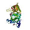
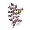
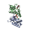
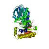
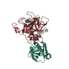
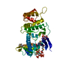
| Title | Crystal Structure of Calcium-Bound Protease Core of Calpain I | ||||||
 Components Components | thiol protease DOMAINS I AND II | ||||||
 Keywords Keywords |  HYDROLASE / PAPAIN-RELATED / CALCIUM-DEPENDENT CYSTEINE PROTEASE / TWO NOVEL COOPERATIVE CALCIUM SITES HYDROLASE / PAPAIN-RELATED / CALCIUM-DEPENDENT CYSTEINE PROTEASE / TWO NOVEL COOPERATIVE CALCIUM SITES | ||||||
| Function / homology |  Function and homology informationcalpain-1 / Degradation of the extracellular matrix / mammary gland involution / positive regulation of leukocyte tethering or rolling / calcium-dependent cysteine-type endopeptidase activity / receptor catabolic process / negative regulation of actin filament polymerization / self proteolysis / cornified envelope / regulation of catalytic activity ...calpain-1 / Degradation of the extracellular matrix / mammary gland involution / positive regulation of leukocyte tethering or rolling / calcium-dependent cysteine-type endopeptidase activity / receptor catabolic process / negative regulation of actin filament polymerization / self proteolysis / cornified envelope / regulation of catalytic activity / positive regulation of vascular permeability / response to arsenic-containing substance / negative regulation of non-canonical NF-kappaB signal transduction / response to angiotensin / Neutrophil degranulation / positive regulation of cardiac muscle cell apoptotic process / protein autoprocessing / cytoskeletal protein binding / protein catabolic process / cellular response to hydrogen peroxide / peptidase activity / lysosome / calcium ion binding / enzyme binding / mitochondrion / proteolysis / membrane / plasma membrane / cytosol / cytoplasm Function and homology informationcalpain-1 / Degradation of the extracellular matrix / mammary gland involution / positive regulation of leukocyte tethering or rolling / calcium-dependent cysteine-type endopeptidase activity / receptor catabolic process / negative regulation of actin filament polymerization / self proteolysis / cornified envelope / regulation of catalytic activity ...calpain-1 / Degradation of the extracellular matrix / mammary gland involution / positive regulation of leukocyte tethering or rolling / calcium-dependent cysteine-type endopeptidase activity / receptor catabolic process / negative regulation of actin filament polymerization / self proteolysis / cornified envelope / regulation of catalytic activity / positive regulation of vascular permeability / response to arsenic-containing substance / negative regulation of non-canonical NF-kappaB signal transduction / response to angiotensin / Neutrophil degranulation / positive regulation of cardiac muscle cell apoptotic process / protein autoprocessing / cytoskeletal protein binding / protein catabolic process / cellular response to hydrogen peroxide / peptidase activity / lysosome / calcium ion binding / enzyme binding / mitochondrion / proteolysis / membrane / plasma membrane / cytosol / cytoplasmSimilarity search - Function | ||||||
| Biological species |  Rattus norvegicus (Norway rat) Rattus norvegicus (Norway rat) | ||||||
| Method | X-RAY DIFFRACTION / MOLECULAR REPLACEMENT / Resolution: 2.07 Å | ||||||
 Authors Authors | Moldoveanu, T. / Hosfield, C.M. / Lim, D. / Elce, J.S. / Jia, Z. / Davies, P.L. | ||||||
 Citation Citation | Journal: Cell(Cambridge,Mass.) / Year: 2002 Title: A Ca(2+) switch aligns the active site of calpain. Authors: Moldoveanu, T. / Hosfield, C.M. / Lim, D. / Elce, J.S. / Jia, Z. / Davies, P.L. | ||||||
| History |
|
- Structure visualization
| Structure viewer | Molecule: MolmilJmol/JSmol |
|---|
- Downloads & links
-Download
| PDBx/mmCIF format | 1kxr.cif.gz | 149 KB | Display | PDBx/mmCIF format |
|---|---|---|---|---|
| PDB format | pdb1kxr.ent.gz | 115.5 KB | Display | PDB format |
| PDBx/mmJSON format | 1kxr.json.gz | Tree view | PDBx/mmJSON format | |
| Others |  Other downloads Other downloads |
-Validation report
| Arichive directory | https://data.pdbj.org/pub/pdb/validation_reports/kx/1kxrftp://data.pdbj.org/pub/pdb/validation_reports/kx/1kxr | HTTPS FTP |
|---|
-Related structure data
| Related structure data |  1df0S S: Starting model for refinement |
|---|---|
| Similar structure data |


-Links
 PDBj
PDBj


- Assembly
| Deposited unit | 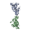
| ||||||||
|---|---|---|---|---|---|---|---|---|---|
| 1 | 
| ||||||||
| 2 | 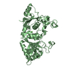
| ||||||||
| Unit cell |
|
-Components
| #1: Protein | Mass: 38788.488 Da / Num. of mol.: 2 / Fragment: RESIDUES 27-356 / Mutation: C115S Source method: isolated from a genetically manipulated source Source: (gene. exp.) Rattus norvegicus (Norway rat) / Gene: CAPN1 / Plasmid: pET24d / Species (production host): Escherichia coli / Production host:  Escherichia coli BL21(DE3) (bacteria) / Strain (production host): BL21-DE3 / References: UniProt: P97571, EC: 3.4.22.17 Escherichia coli BL21(DE3) (bacteria) / Strain (production host): BL21-DE3 / References: UniProt: P97571, EC: 3.4.22.17#2: Chemical | ChemComp-CA /   Mass: 40.078 Da / Num. of mol.: 4 / Source method: obtained synthetically / Formula: Ca Mass: 40.078 Da / Num. of mol.: 4 / Source method: obtained synthetically / Formula: Ca#3: Water | ChemComp-HOH / | Water Mass: 18.015 Da / Num. of mol.: 359 / Source method: isolated from a natural source / Formula: H2O Mass: 18.015 Da / Num. of mol.: 359 / Source method: isolated from a natural source / Formula: H2O |
|---|
-Experimental details
-Experiment
| Experiment | Method: X-RAY DIFFRACTION / Number of used crystals: 1 |
|---|
- Sample preparation
| Crystal | Density Matthews: 2.48 Å3/Da / Density % sol: 50.41 % | |||||||||||||||||||||||||||||||||||||||||||||||||
|---|---|---|---|---|---|---|---|---|---|---|---|---|---|---|---|---|---|---|---|---|---|---|---|---|---|---|---|---|---|---|---|---|---|---|---|---|---|---|---|---|---|---|---|---|---|---|---|---|---|---|
| Crystal grow | Temperature: 298 K / Method: vapor diffusion, hanging drop / pH: 6 Details: sodium chloride, calcium chloride, MES, PEG 6000, pH 6.0, VAPOR DIFFUSION, HANGING DROP, temperature 298.0K | |||||||||||||||||||||||||||||||||||||||||||||||||
| Crystal grow | *PLUS | |||||||||||||||||||||||||||||||||||||||||||||||||
| Components of the solutions | *PLUS
|
-Data collection
| Diffraction | Mean temperature: 100 K |
|---|---|
| Diffraction source | Source: ROTATING ANODE / Type: RIGAKU RU200 / Wavelength: 1.5418 Å |
| Detector | Type: MARRESEARCH / Detector: IMAGE PLATE / Date: Mar 5, 2001 / Details: mirrors |
| Radiation | Monochromator: YALE MIRRORS / Protocol: SINGLE WAVELENGTH / Monochromatic (M) / Laue (L): M / Scattering type: x-ray |
| Radiation wavelength | Wavelength: 1.5418 Å / Relative weight: 1 |
| Reflection | Resolution: 2.07→30 Å / Num. obs: 43051 / % possible obs: 92 % / Observed criterion σ(F): 2 / Observed criterion σ(I): 2 / Redundancy: 8 % / Rmerge(I) obs: 0.091 / Net I/σ(I): 11.3 |
| Reflection shell | Resolution: 2.07→2.14 Å / Redundancy: 5.4 % / Rmerge(I) obs: 0.39 / Mean I/σ(I) obs: 2.8 / Num. unique all: 4388 / Rsym value: 0.368 / % possible all: 94.6 |
| Reflection | *PLUS % possible obs: 92 % / Num. measured all: 119331 |
| Reflection shell | *PLUS Rmerge(I) obs: 0.14 |
- Processing
| Software |
| ||||||||||||||||||||||||||||||||||||||||||
|---|---|---|---|---|---|---|---|---|---|---|---|---|---|---|---|---|---|---|---|---|---|---|---|---|---|---|---|---|---|---|---|---|---|---|---|---|---|---|---|---|---|---|---|
| Refinement | Method to determine structure: MOLECULAR REPLACEMENT Starting model: pdb entry 1DF0 Resolution: 2.07→30 Å / Cross valid method: THROUGHOUT / σ(F): 0 / Stereochemistry target values: Engh & Huber
| ||||||||||||||||||||||||||||||||||||||||||
| Refinement step | Cycle: LAST / Resolution: 2.07→30 Å
| ||||||||||||||||||||||||||||||||||||||||||
| Refine LS restraints |
| ||||||||||||||||||||||||||||||||||||||||||
| LS refinement shell |
| ||||||||||||||||||||||||||||||||||||||||||
| Refinement | *PLUS % reflection Rfree: 5 % / Rfactor obs: 0.218 / Rfactor Rfree: 0.257 | ||||||||||||||||||||||||||||||||||||||||||
| Solvent computation | *PLUS | ||||||||||||||||||||||||||||||||||||||||||
| Displacement parameters | *PLUS | ||||||||||||||||||||||||||||||||||||||||||
| Refine LS restraints | *PLUS
| ||||||||||||||||||||||||||||||||||||||||||
| LS refinement shell | *PLUS Rfactor obs: 0.2432 |
