 Movie
Movie Controller


+ Open data

- Basic information
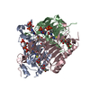
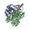
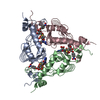
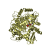
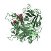
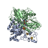
| Entry | Database: PDB / ID: 1krr | ||||||
|---|---|---|---|---|---|---|---|
| Title | Galactoside Acetyltransferase in Complex with Acetyl-Coenzyme A | ||||||
 Components Components | GALACTOSIDE O-ACETYLTRANSFERASE Galactoside acetyltransferase Galactoside acetyltransferase | ||||||
 Keywords Keywords | TRANSFERASE / Left-Handed Parallel Beta Helix | ||||||
| Function / homology |  Function and homology informationgalactoside O-acetyltransferase / galactoside O-acetyltransferase activity / lactose biosynthetic process / identical protein binding / cytoplasm Function and homology informationgalactoside O-acetyltransferase / galactoside O-acetyltransferase activity / lactose biosynthetic process / identical protein binding / cytoplasmSimilarity search - Function | ||||||
| Biological species |  Escherichia coli (E. coli) Escherichia coli (E. coli) | ||||||
| Method | X-RAY DIFFRACTION / MIR / Resolution: 2.5 Å | ||||||
 Authors Authors | Wang, X.-G. / Olsen, L.R. / Roderick, S.L. | ||||||
 Citation Citation | Journal: Structure / Year: 2002 Title: Structure of the lac operon galactoside acetyltransferase. Authors: Wang, X.G. / Olsen, L.R. / Roderick, S.L. | ||||||
| History |
|
- Structure visualization
| Structure viewer | Molecule: MolmilJmol/JSmol |
|---|
- Downloads & links
-Download
| PDBx/mmCIF format | 1krr.cif.gz | 131.3 KB | Display | PDBx/mmCIF format |
|---|---|---|---|---|
| PDB format | pdb1krr.ent.gz | 104.2 KB | Display | PDB format |
| PDBx/mmJSON format | 1krr.json.gz | Tree view | PDBx/mmJSON format | |
| Others |  Other downloads Other downloads |
-Validation report
| Arichive directory | https://data.pdbj.org/pub/pdb/validation_reports/kr/1krrftp://data.pdbj.org/pub/pdb/validation_reports/kr/1krr | HTTPS FTP |
|---|
-Related structure data


-Links
 PDBj
PDBj


- Assembly
| Deposited unit | 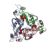
| ||||||||
|---|---|---|---|---|---|---|---|---|---|
| 1 |
| ||||||||
| Unit cell |
| ||||||||
| Details | The biological assembly is a trimer. |
-Components
| #1: Protein | Galactoside acetyltransferase / THIOGALACTOSIDE ACETYLTRANSFERASE Mass: 22826.998 Da / Num. of mol.: 3 Source method: isolated from a genetically manipulated source Source: (gene. exp.) Escherichia coli (E. coli) / Gene: lacA / Plasmid: ptac-85 / Production host: Escherichia coli (E. coli) / Strain (production host): TG1References: UniProt: P07464, galactoside O-acetyltransferase#2: Chemical | Acetyl-CoA  Mass: 809.571 Da / Num. of mol.: 3 / Source method: obtained synthetically / Formula: C23H38N7O17P3S Mass: 809.571 Da / Num. of mol.: 3 / Source method: obtained synthetically / Formula: C23H38N7O17P3S#3: Water | ChemComp-HOH / | Water Mass: 18.015 Da / Num. of mol.: 113 / Source method: isolated from a natural source / Formula: H2O Mass: 18.015 Da / Num. of mol.: 113 / Source method: isolated from a natural source / Formula: H2O |
|---|
-Experimental details
-Experiment
| Experiment | Method: X-RAY DIFFRACTION / Number of used crystals: 1 |
|---|
- Sample preparation
| Crystal | Density Matthews: 2.65 Å3/Da / Density % sol: 53.61 % | ||||||||||||||||||||||||||||||||||||||||||||||||
|---|---|---|---|---|---|---|---|---|---|---|---|---|---|---|---|---|---|---|---|---|---|---|---|---|---|---|---|---|---|---|---|---|---|---|---|---|---|---|---|---|---|---|---|---|---|---|---|---|---|
| Crystal grow | Temperature: 298 K / Method: vapor diffusion, hanging drop / pH: 7.8 Details: Tris-HCl, beta-mercaptoethanol, ammonium sulfate, HEPES, tartaric acid, acetyl-CoA, pH 7.8, VAPOR DIFFUSION, HANGING DROP, temperature 298K | ||||||||||||||||||||||||||||||||||||||||||||||||
| Crystal grow | *PLUS Details: Wang, X.G., (1999) Acta Crystallogr., D55, 1955. | ||||||||||||||||||||||||||||||||||||||||||||||||
| Components of the solutions | *PLUS
|
-Data collection
| Diffraction | Mean temperature: 298 K |
|---|---|
| Diffraction source | Source: ROTATING ANODE / Type: RIGAKU RU200 / Wavelength: 1.5418 Å |
| Detector | Type: SIEMENS HI-STAR / Detector: AREA DETECTOR / Date: Dec 2, 1998 / Details: mirrors |
| Radiation | Monochromator: Confocal / Protocol: SINGLE WAVELENGTH / Monochromatic (M) / Laue (L): M / Scattering type: x-ray |
| Radiation wavelength | Wavelength: 1.5418 Å / Relative weight: 1 |
| Reflection | Resolution: 2.5→26 Å / Num. all: 23094 / Num. obs: 23094 / % possible obs: 90.1 % / Observed criterion σ(F): 0 / Observed criterion σ(I): 0 / Redundancy: 4 % / Biso Wilson estimate: 22.6 Å2 / Rmerge(I) obs: 0.089 |
| Reflection shell | Resolution: 2.5→2.6 Å / Rmerge(I) obs: 0.298 / % possible all: 68.8 |
| Reflection | *PLUS Num. measured all: 93176 / Rmerge(I) obs: 0.089 |
| Reflection shell | *PLUS Highest resolution: 2.5 Å / Lowest resolution: 2.6 Å / % possible obs: 68.8 % / Rmerge(I) obs: 0.298 |
- Processing
| Software |
| ||||||||||||||||||||||||||||||||||||
|---|---|---|---|---|---|---|---|---|---|---|---|---|---|---|---|---|---|---|---|---|---|---|---|---|---|---|---|---|---|---|---|---|---|---|---|---|---|
| Refinement | Method to determine structure: MIR / Resolution: 2.5→26 Å / Rfactor Rfree error: 0.007 / Data cutoff high absF: 10000000 / Data cutoff low absF: 0.001 / Isotropic thermal model: RESTRAINED / Cross valid method: THROUGHOUT / σ(F): 0 / Stereochemistry target values: Engh & Huber
| ||||||||||||||||||||||||||||||||||||
| Displacement parameters | Biso mean: 21.8 Å2
| ||||||||||||||||||||||||||||||||||||
| Refine analyze |
| ||||||||||||||||||||||||||||||||||||
| Refinement step | Cycle: LAST / Resolution: 2.5→26 Å
| ||||||||||||||||||||||||||||||||||||
| Refine LS restraints |
| ||||||||||||||||||||||||||||||||||||
| LS refinement shell | Resolution: 2.5→2.59 Å / Rfactor Rfree error: 0.037 / Total num. of bins used: 10
| ||||||||||||||||||||||||||||||||||||
| Xplor file |
| ||||||||||||||||||||||||||||||||||||
| Refinement | *PLUS Rfactor all: 0.196 / Rfactor obs: 0.193 / Rfactor Rfree: 0.248 / Rfactor Rwork: 0.193 | ||||||||||||||||||||||||||||||||||||
| Solvent computation | *PLUS | ||||||||||||||||||||||||||||||||||||
| Displacement parameters | *PLUS | ||||||||||||||||||||||||||||||||||||
| Refine LS restraints | *PLUS
| ||||||||||||||||||||||||||||||||||||
| LS refinement shell | *PLUS Highest resolution: 2.5 Å / Lowest resolution: 2.6 Å / Rfactor Rfree: 0.332 / Rfactor Rwork: 0.252 / Rfactor obs: 0.252 |
