 Movie
Movie Controller

[English] 日本語
 Yorodumi
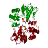
- PDB-1kmz: MOLECULAR BASIS OF MITOMYCIN C RESICTANCE IN STREPTOMYCES: CRYSTA... -
+ Open data

- Basic information
| Entry | Database: PDB / ID: 1kmz | ||||||
|---|---|---|---|---|---|---|---|
| Title | MOLECULAR BASIS OF MITOMYCIN C RESICTANCE IN STREPTOMYCES: CRYSTAL STRUCTURES OF THE MRD PROTEIN WITH AND WITHOUT A DRUG DERIVATIVE | ||||||
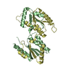
 Components Components | mitomycin-binding protein | ||||||
 Keywords Keywords |  ANTIMICROBIAL PROTEIN / Mitomycin C / antibiotic resistance / SAD / anomalous diffraction / domain swapping / p-staking ANTIMICROBIAL PROTEIN / Mitomycin C / antibiotic resistance / SAD / anomalous diffraction / domain swapping / p-staking | ||||||
| Function / homology |  Function and homology information Function and homology information2,3-Dihydroxybiphenyl 1,2-Dioxygenase, domain 1 / 2,3-Dihydroxybiphenyl 1,2-Dioxygenase; domain 1 / Glyoxalase/fosfomycin resistance/dioxygenase domain / Glyoxalase/Bleomycin resistance protein/Dioxygenase superfamily / Vicinal oxygen chelate (VOC) domain / Vicinal oxygen chelate (VOC) domain profile. / Glyoxalase/Bleomycin resistance protein/Dihydroxybiphenyl dioxygenase / Roll / Alpha Beta Similarity search - Domain/homology | ||||||
| Biological species |  Streptomyces lavendulae (bacteria) Streptomyces lavendulae (bacteria) | ||||||
| Method | X-RAY DIFFRACTION / SYNCHROTRON / Resolution: 1.5 Å | ||||||
 Authors Authors | Martin, T.W. / Dauter, Z. / Devedjiev, Y. / Sheffield, P. / Jelen, F. / He, M. / Sherman, D. / Otlewski, J. / Derewenda, Z.S. / Derewenda, U. | ||||||
 Citation Citation | Journal: Structure / Year: 2002 Title: Molecular basis of mitomycin C resistance in streptomyces: structure and function of the MRD protein. Authors: Martin, T.W. / Dauter, Z. / Devedjiev, Y. / Sheffield, P. / Jelen, F. / He, M. / Sherman, D.H. / Otlewski, J. / Derewenda, Z.S. / Derewenda, U. | ||||||
| History |
|
- Structure visualization
| Structure viewer | Molecule: MolmilJmol/JSmol |
|---|
- Downloads & links
-Download
| PDBx/mmCIF format | 1kmz.cif.gz | 70.7 KB | Display | PDBx/mmCIF format |
|---|---|---|---|---|
| PDB format | pdb1kmz.ent.gz | 51.4 KB | Display | PDB format |
| PDBx/mmJSON format | 1kmz.json.gz | Tree view | PDBx/mmJSON format | |
| Others |  Other downloads Other downloads |
-Validation report
| Arichive directory | https://data.pdbj.org/pub/pdb/validation_reports/km/1kmzftp://data.pdbj.org/pub/pdb/validation_reports/km/1kmz | HTTPS FTP |
|---|
-Related structure data
| Related structure data | 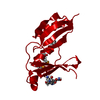 1kllSC S: Starting model for refinement C: citing same article ( |
|---|---|
| Similar structure data |


-Links
 PDBj
PDBj- Assembly
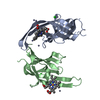
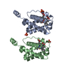
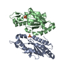
| Deposited unit | 
| ||||||||||
|---|---|---|---|---|---|---|---|---|---|---|---|
| 1 | 
| ||||||||||
| 2 | 
| ||||||||||
| Unit cell |
| ||||||||||
| Details | The second part of the biological assembly is generated by the crystallographyc two fold axis |
-Components
| #1: Protein | Mass: 14395.127 Da / Num. of mol.: 1 Source method: isolated from a genetically manipulated source Source: (gene. exp.) Streptomyces lavendulae (bacteria) / Production host: Escherichia coli (E. coli) / Strain (production host): BL221(DE3) / References: UniProt: O05205 |
|---|---|
| #2: Water | ChemComp-HOH / Water Mass: 18.015 Da / Num. of mol.: 170 / Source method: isolated from a natural source / Formula: H2O Mass: 18.015 Da / Num. of mol.: 170 / Source method: isolated from a natural source / Formula: H2O |
-Experimental details
-Experiment
| Experiment | Method: X-RAY DIFFRACTION / Number of used crystals: 1 |
|---|
- Sample preparation
| Crystal | Density Matthews: 1.7 Å3/Da / Density % sol: 37.62 % | ||||||||||||||||||||||||
|---|---|---|---|---|---|---|---|---|---|---|---|---|---|---|---|---|---|---|---|---|---|---|---|---|---|
| Crystal grow | Temperature: 298 K / Method: vapor diffusion / pH: 6 Details: ammonium sulfate, MES buffer, beta-octylglucoside, pH 6.0, VAPOR DIFFUSION at 298K, temperature 298.0K | ||||||||||||||||||||||||
| Crystal grow | *PLUS Method: unknown | ||||||||||||||||||||||||
| Components of the solutions | *PLUS
|
-Data collection
| Diffraction | Mean temperature: 100 K |
|---|---|
| Diffraction source | Source: SYNCHROTRON / Site: NSLS  / Beamline: X9B / Wavelength: 0.97 / Beamline: X9B / Wavelength: 0.97 |
| Detector | Type: ADSC QUANTUM 4 / Detector: CCD / Date: Jul 24, 2000 / Details: MIRRORS |
| Radiation | Monochromator: Si 111 / Protocol: SINGLE WAVELENGTH / Monochromatic (M) / Laue (L): M / Scattering type: x-ray |
| Radiation wavelength | Wavelength: 0.97 Å / Relative weight: 1 |
| Reflection | Resolution: 1.5→30 Å / Num. obs: 17820 / % possible obs: 99.9 % / Redundancy: 4.15 % / Biso Wilson estimate: 18.6 Å2 / Rmerge(I) obs: 0.034 |
| Reflection shell | Resolution: 1.5→1.58 Å / Redundancy: 4.1 % / Rmerge(I) obs: 0.33 / % possible all: 100 |
| Reflection | *PLUS Highest resolution: 1.5 Å / Num. measured all: 74125 / Rmerge(I) obs: 0.034 |
| Reflection shell | *PLUS % possible obs: 99.4 % / Rmerge(I) obs: 0.335 / Mean I/σ(I) obs: 3.6 |
- Processing
| Software |
| ||||||||||||||||||||
|---|---|---|---|---|---|---|---|---|---|---|---|---|---|---|---|---|---|---|---|---|---|
| Refinement | Starting model: PDB ENTRY 1KLL Resolution: 1.5→10 Å / Cross valid method: FREE R / σ(F): 1 / Stereochemistry target values: ENGH & HUBER
| ||||||||||||||||||||
| Refine analyze | Luzzati coordinate error obs: 0.15 Å / Num. disordered residues: 5 | ||||||||||||||||||||
| Refinement step | Cycle: LAST / Resolution: 1.5→10 Å
| ||||||||||||||||||||
| Refine LS restraints |
| ||||||||||||||||||||
| Software | *PLUS Name: SHELXL / Version: 97 / Classification: refinement | ||||||||||||||||||||
| Refinement | *PLUS Rfactor obs: 0.163 / Rfactor Rfree: 0.234 / Rfactor Rwork: 0.168 | ||||||||||||||||||||
| Solvent computation | *PLUS | ||||||||||||||||||||
| Displacement parameters | *PLUS | ||||||||||||||||||||
| Refine LS restraints | *PLUS
|
