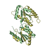
Entry Database : PDB / ID : 5zyoTitle Crystal Structure of domain-swapped Circular-Permuted YbeA (CP74) from Escherichia coli Ribosomal RNA large subunit methyltransferase H Keywords / / / / Function / homology Biological species Escherichia coli K-12 (bacteria)Method / / / Resolution : 1.75 Å Authors Ko, K.T. / Huang, K.F. / Lyu, P.C. / Hsu, S.T.D. Funding support Organization Grant number Country Ministry of Science and Technology (Taiwan) 106-2113-M-001-004 Ministry of Science and Technology (Taiwan) 107-2628-M-001-005-MY3 Ministry of Science and Technology (Taiwan) 106-2311-B-007-004-MY3 Academia Sinica (Taiwan)
Journal : Structure / Year : 2019Title : Untying a Knotted SPOUT RNA Methyltransferase by Circular Permutation Results in a Domain-Swapped Dimer.Authors : Ko, K.T. / Hu, I.C. / Huang, K.F. / Lyu, P.C. / Hsu, S.D. History Deposition May 26, 2018 Deposition site / Processing site Revision 1.0 May 29, 2019 Provider / Type Revision 1.1 Jun 5, 2019 Group / Database references / Category / citation_authorItem _citation.pdbx_database_id_PubMed / _citation.title ... _citation.pdbx_database_id_PubMed / _citation.title / _citation_author.identifier_ORCID / _citation_author.name Revision 1.2 Aug 21, 2019 Group / Database references / Category Item / _citation.page_first / _citation.page_lastRevision 1.3 Mar 27, 2024 Group / Database references / Category / chem_comp_bond / database_2Item / _database_2.pdbx_database_accession
Show all Show less
 Movie
Movie Controller

 Yorodumi
 Open data
 Basic information
 Components
 Keywords
 Function and homology information
 Authors
 Taiwan, 4items
  Citation
 Structure visualization
 Downloads & links
 Other downloads
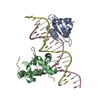


 PDBj
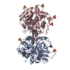
PDBj Assembly

 Mass: 18.015 Da / Num. of mol.: 351 / Source method: isolated from a natural source / Formula: H2O
 Sample preparation
 Processing