 Movie
Movie Controller

[English] 日本語
 Yorodumi
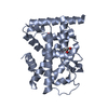
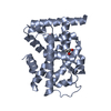
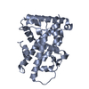
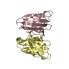
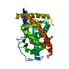
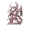
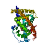
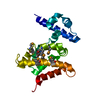
- PDB-1jv6: BACTERIORHODOPSIN D85S/F219L DOUBLE MUTANT AT 2.00 ANGSTROM RESOLUTION -
+ Open data

- Basic information
| Entry | Database: PDB / ID: 1jv6 | ||||||
|---|---|---|---|---|---|---|---|
| Title | BACTERIORHODOPSIN D85S/F219L DOUBLE MUTANT AT 2.00 ANGSTROM RESOLUTION | ||||||
 Components Components | Bacteriorhodopsin | ||||||
 Keywords Keywords | ION TRANSPORT / PHOTORECEPTOR / HALOARCHAEA / 7-TRANSMEMBRANE / F219L MUTANT / CUBIC LIPID PHASE | ||||||
| Function / homology |  Function and homology informationphotoreceptor activity / phototransduction / proton transmembrane transport / monoatomic ion channel activity / plasma membrane Function and homology informationphotoreceptor activity / phototransduction / proton transmembrane transport / monoatomic ion channel activity / plasma membraneSimilarity search - Function | ||||||
| Biological species |  Halobacterium salinarum (Halophile) Halobacterium salinarum (Halophile) | ||||||
| Method | X-RAY DIFFRACTION / SYNCHROTRON / MOLECULAR REPLACEMENT / Resolution: 2 Å | ||||||
 Authors Authors | Rouhani, S. / Cartailler, J.-P. / Facciotti, M.T. / Walian, P. / Needleman, R. / Lanyi, J.K. / Glaeser, R.M. / Luecke, H. | ||||||
 Citation Citation | Journal: J.Mol.Biol. / Year: 2001 Title: Crystal structure of the D85S mutant of bacteriorhodopsin: model of an O-like photocycle intermediate. Authors: Rouhani, S. / Cartailler, J.P. / Facciotti, M.T. / Walian, P. / Needleman, R. / Lanyi, J.K. / Glaeser, R.M. / Luecke, H. | ||||||
| History |
|
- Structure visualization
| Structure viewer | Molecule: MolmilJmol/JSmol |
|---|
- Downloads & links
-Download
| PDBx/mmCIF format | 1jv6.cif.gz | 60.8 KB | Display | PDBx/mmCIF format |
|---|---|---|---|---|
| PDB format | pdb1jv6.ent.gz | 41.6 KB | Display | PDB format |
| PDBx/mmJSON format | 1jv6.json.gz | Tree view | PDBx/mmJSON format | |
| Others |  Other downloads Other downloads |
-Validation report
| Arichive directory | https://data.pdbj.org/pub/pdb/validation_reports/jv/1jv6ftp://data.pdbj.org/pub/pdb/validation_reports/jv/1jv6 | HTTPS FTP |
|---|
-Related structure data
| Related structure data | 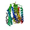 1jv7SC S: Starting model for refinement C: citing same article ( |
|---|---|
| Similar structure data |


-Links
 PDBj
PDBj


- Assembly
| Deposited unit | 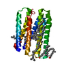
| ||||||||
|---|---|---|---|---|---|---|---|---|---|
| 1 |
| ||||||||
| Unit cell |
|
-Components
| #1: Protein | / BR Mass: 26867.475 Da / Num. of mol.: 1 / Mutation: D85S,F219L Source method: isolated from a genetically manipulated source Source: (gene. exp.) Halobacterium salinarum (Halophile) / Gene: Bop / Plasmid: pNov-r / Production host: Halobacterium salinarum (Halophile) / References: UniProt: P02945 | ||
|---|---|---|---|
| #2: Chemical | ChemComp-RET / Retinal  Mass: 284.436 Da / Num. of mol.: 1 / Source method: obtained synthetically / Formula: C20H28O Mass: 284.436 Da / Num. of mol.: 1 / Source method: obtained synthetically / Formula: C20H28O | ||
| #3: Chemical | ChemComp-LI1 /   Mass: 639.130 Da / Num. of mol.: 8 / Source method: obtained synthetically / Formula: C42H86O3 Mass: 639.130 Da / Num. of mol.: 8 / Source method: obtained synthetically / Formula: C42H86O3#4: Water | ChemComp-HOH / | Water Mass: 18.015 Da / Num. of mol.: 34 / Source method: isolated from a natural source / Formula: H2O Mass: 18.015 Da / Num. of mol.: 34 / Source method: isolated from a natural source / Formula: H2O |
-Experimental details
-Experiment
| Experiment | Method: X-RAY DIFFRACTION / Number of used crystals: 1 |
|---|
- Sample preparation
| Crystal | Density Matthews: 2.6 Å3/Da / Density % sol: 52.1 % | |||||||||||||||||||||||||
|---|---|---|---|---|---|---|---|---|---|---|---|---|---|---|---|---|---|---|---|---|---|---|---|---|---|---|
| Crystal grow | Temperature: 293 K / Method: cubic lipid phase / pH: 5.6 Details: monoolein, octyl-beta-D-glucopyranoside, pH 5.6, cubic lipid phase, temperature 293K | |||||||||||||||||||||||||
| Crystal grow | *PLUS Temperature: 20 ℃Details: Landau, E.M., (1996) Proc.Natl.Acad.Sci.USA., 93, 14532. | |||||||||||||||||||||||||
| Components of the solutions | *PLUS
|
-Data collection
| Diffraction |
| |||||||||
|---|---|---|---|---|---|---|---|---|---|---|
| Diffraction source | Source: SYNCHROTRON / Site: ALS  / Beamline: 5.0.2 / Wavelength: 1 Å / Beamline: 5.0.2 / Wavelength: 1 Å | |||||||||
| Detector | Type: ADSC QUANTUM / Detector: CCD / Date: Apr 7, 1999 | |||||||||
| Radiation | Monochromator: double crystal monochromator / Protocol: SINGLE WAVELENGTH / Monochromatic (M) / Laue (L): M / Scattering type: x-ray | |||||||||
| Radiation wavelength | Wavelength: 1 Å / Relative weight: 1 | |||||||||
| Reflection | Resolution: 2→30 Å / Num. all: 18717 / Num. obs: 14934 / % possible obs: 79.8 % / Observed criterion σ(F): 1 / Observed criterion σ(I): 1 / Redundancy: 8.1 % / Rsym value: 0.077 / Net I/σ(I): 14.4 | |||||||||
| Reflection shell | Resolution: 2→2.07 Å / Mean I/σ(I) obs: 2.6 / Num. unique all: 769 / Rsym value: 0.558 / % possible all: 42 | |||||||||
| Reflection | *PLUS Highest resolution: 2 Å / Lowest resolution: 99 Å / Num. measured all: 152520 / Rmerge(I) obs: 0.077 | |||||||||
| Reflection shell | *PLUS % possible obs: 70.8 % / Rmerge(I) obs: 0.558 |
- Processing
| Software |
| |||||||||||||||||||||||||
|---|---|---|---|---|---|---|---|---|---|---|---|---|---|---|---|---|---|---|---|---|---|---|---|---|---|---|
| Refinement | Method to determine structure: MOLECULAR REPLACEMENT Starting model: PDB ENTRY 1JV7 (Bacteriorhodopsin D85S coordinates, without diether lipids nor water) Resolution: 2→12 Å / Isotropic thermal model: isotropic / Cross valid method: THROUGHOUT / σ(F): 1 / σ(I): 1 / Stereochemistry target values: Engh & Huber Details: Intensities (I) and not amplitude (F) was used in refinement.
| |||||||||||||||||||||||||
| Refinement step | Cycle: LAST / Resolution: 2→12 Å
| |||||||||||||||||||||||||
| Refine LS restraints |
| |||||||||||||||||||||||||
| Xplor file | Serial no: 1 / Param file: protein_rep.param / Topol file: protein.top | |||||||||||||||||||||||||
| Software | *PLUS Name: CNS / Version: 1 / Classification: refinement | |||||||||||||||||||||||||
| Refinement | *PLUS Lowest resolution: 12 Å / σ(F): 1 / % reflection Rfree: 5 % / Rfactor obs: 0.222 / Rfactor Rfree: 0.24 | |||||||||||||||||||||||||
| Solvent computation | *PLUS | |||||||||||||||||||||||||
| Displacement parameters | *PLUS |
