 Movie
Movie Controller

[English] 日本語
 Yorodumi
- PDB-1fcz: ISOTYPE SELECTIVITY OF THE HUMAN RETINOIC ACID NUCLEAR RECEPTOR H... -
+ Open data

- Basic information
| Entry | Database: PDB / ID: 1fcz | ||||||
|---|---|---|---|---|---|---|---|
| Title | ISOTYPE SELECTIVITY OF THE HUMAN RETINOIC ACID NUCLEAR RECEPTOR HRAR: THE COMPLEX WITH THE PANAGONIST RETINOID BMS181156 | ||||||
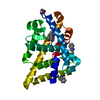
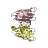
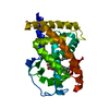
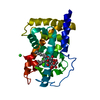
 Components Components | RETINOIC ACID RECEPTOR GAMMA-1 | ||||||
 Keywords Keywords | GENE REGULATION / isotype selectivity / retinoid ligand complexes / drug design / antiparallel alpha-helical sandwich fold / Structural Proteomics in Europe / SPINE / Structural Genomics | ||||||
| Function / homology |  Function and homology information Function and homology informationHarderian gland development / regulation of myeloid cell differentiation / growth plate cartilage chondrocyte growth / trachea cartilage development / embryonic eye morphogenesis / embryonic camera-type eye development / glandular epithelial cell development / cellular response to corticotropin-releasing hormone stimulus / nuclear glucocorticoid receptor binding / prostate gland epithelium morphogenesis ...Harderian gland development / regulation of myeloid cell differentiation / growth plate cartilage chondrocyte growth / trachea cartilage development / embryonic eye morphogenesis / embryonic camera-type eye development / glandular epithelial cell development / cellular response to corticotropin-releasing hormone stimulus / nuclear glucocorticoid receptor binding / prostate gland epithelium morphogenesis / positive regulation of programmed cell death / negative regulation of chondrocyte differentiation / embryonic hindlimb morphogenesis / anterior/posterior pattern specification / regulation of myelination / Signaling by Retinoic Acid / regulation of cell size / face development / retinoic acid receptor signaling pathway / canonical Wnt signaling pathway / nuclear retinoid X receptor binding / negative regulation of stem cell proliferation / cellular response to retinoic acid / response to retinoic acid / hormone-mediated signaling pathway / cellular response to leukemia inhibitory factor / stem cell proliferation / neural tube closure / multicellular organism growth / Activation of anterior HOX genes in hindbrain development during early embryogenesis / Nuclear Receptor transcription pathway / nuclear receptor activity / sequence-specific double-stranded DNA binding / transcription regulator complex / cell differentiation / DNA-binding transcription factor activity, RNA polymerase II-specific / positive regulation of apoptotic process / RNA polymerase II cis-regulatory region sequence-specific DNA binding / DNA-binding transcription factor activity / negative regulation of cell population proliferation / apoptotic process / chromatin binding / chromatin / positive regulation of cell population proliferation / positive regulation of gene expression / negative regulation of transcription by RNA polymerase II / positive regulation of transcription by RNA polymerase II / DNA binding / zinc ion binding / nucleoplasm / membrane / nucleus / cytoplasmSimilarity search - Function | ||||||
| Biological species |  Homo sapiens (human) Homo sapiens (human) | ||||||
| Method | X-RAY DIFFRACTION / SYNCHROTRON / MOLECULAR REPLACEMENT / Resolution: 1.38 Å | ||||||
 Authors Authors | Klaholz, B.P. / Mitschler, A. / Moras, D. / Structural Proteomics in Europe (SPINE) | ||||||
 Citation Citation | Journal: J.Mol.Biol. / Year: 2000 Title: Structural basis for isotype selectivity of the human retinoic acid nuclear receptor. Authors: Klaholz, B.P. / Mitschler, A. / Moras, D. #1: Journal: Proc.Natl.Acad.Sci.USA / Year: 2000Title: Enantiomer discrimination illustrated by high-resolution crystal structures of the human nuclear receptor hRARgamma. Authors: Klaholz, B.P. / Mitschler, A. / Belema, M. / Zusi, C. / Moras, D. #2: Journal: Nat.Struct.Biol. / Year: 1998Title: Conformational adaptation of agonists to the human nuclear receptor hRARgamma. Authors: Klaholz, B.P. / Renaud, J.-P. / Mitschler, A. / Zusi, C. / Chambon, P. / Gronemeyer, H. / Moras, D. #3: Journal: Nature / Year: 1995Title: Crystal structure of the RARgamma ligand-binding domain bound to all-trans retinoic acid. Authors: Renaud, J.-P. / Rochel, N. / Ruff, M. / Vivat, V. / Chambon, P. / Gronemeyer, H. / Moras, D. | ||||||
| History |
|
- Structure visualization
| Structure viewer | Molecule: MolmilJmol/JSmol |
|---|
- Downloads & links
-Download
| PDBx/mmCIF format | 1fcz.cif.gz | 72.2 KB | Display | PDBx/mmCIF format |
|---|---|---|---|---|
| PDB format | pdb1fcz.ent.gz | 51.3 KB | Display | PDB format |
| PDBx/mmJSON format | 1fcz.json.gz | Tree view | PDBx/mmJSON format | |
| Others |  Other downloads Other downloads |
-Validation report
| Arichive directory | https://data.pdbj.org/pub/pdb/validation_reports/fc/1fczftp://data.pdbj.org/pub/pdb/validation_reports/fc/1fcz | HTTPS FTP |
|---|
-Related structure data
| Related structure data |  1fcxC  1fcySC S: Starting model for refinement C: citing same article ( |
|---|---|
| Similar structure data | |
| Other databases |


-Links
 PDBj
PDBj
- Assembly
| Deposited unit | 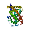
| ||||||||||
|---|---|---|---|---|---|---|---|---|---|---|---|
| 1 |
| ||||||||||
| Unit cell |
|
-Components
| #1: Protein | / RAR-GAMMA-1 Mass: 26535.070 Da / Num. of mol.: 1 / Fragment: LIGAND BINDING DOMAIN Source method: isolated from a genetically manipulated source Details: COMPLEXED WITH BMS181156, RESIDUE 156 / Source: (gene. exp.) Homo sapiens (human) / Plasmid: PET-15B / Production host:  Escherichia coli (E. coli) / References: UniProt: P13631 Escherichia coli (E. coli) / References: UniProt: P13631 |
|---|---|
| #2: Chemical | ChemComp-156 /   Mass: 362.461 Da / Num. of mol.: 1 / Source method: obtained synthetically / Formula: C24H26O3 Mass: 362.461 Da / Num. of mol.: 1 / Source method: obtained synthetically / Formula: C24H26O3 |
| #3: Sugar | ChemComp-LMU / 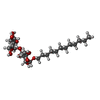  Type: D-saccharide / Mass: 510.615 Da / Num. of mol.: 1 / Source method: obtained synthetically / Formula: C24H46O11 / Comment: detergent*YM Type: D-saccharide / Mass: 510.615 Da / Num. of mol.: 1 / Source method: obtained synthetically / Formula: C24H46O11 / Comment: detergent*YM |
| #4: Water | ChemComp-HOH / Water Mass: 18.015 Da / Num. of mol.: 305 / Source method: isolated from a natural source / Formula: H2O Mass: 18.015 Da / Num. of mol.: 305 / Source method: isolated from a natural source / Formula: H2O |
-Experimental details
-Experiment
| Experiment | Method: X-RAY DIFFRACTION / Number of used crystals: 1 |
|---|
- Sample preparation
| Crystal | Density Matthews: 2.27 Å3/Da / Density % sol: 45.9 % | ||||||||||||||||||||||||||||||||||||||||||||||||||||||||||||
|---|---|---|---|---|---|---|---|---|---|---|---|---|---|---|---|---|---|---|---|---|---|---|---|---|---|---|---|---|---|---|---|---|---|---|---|---|---|---|---|---|---|---|---|---|---|---|---|---|---|---|---|---|---|---|---|---|---|---|---|---|---|
| Crystal grow | Temperature: 290 K / Method: vapor diffusion, hanging drop / pH: 7 Details: sodium acetate, pH 7.0, VAPOR DIFFUSION, HANGING DROP at 290K | ||||||||||||||||||||||||||||||||||||||||||||||||||||||||||||
| Crystal grow | *PLUS Temperature: 17 ℃ | ||||||||||||||||||||||||||||||||||||||||||||||||||||||||||||
| Components of the solutions | *PLUS
|
-Data collection
| Diffraction | Mean temperature: 100 K |
|---|---|
| Diffraction source | Source: SYNCHROTRON / Site: EMBL/DESY, HAMBURG  / Beamline: BW7B / Wavelength: 0.8345 / Beamline: BW7B / Wavelength: 0.8345 |
| Detector | Type: MARRESEARCH / Detector: IMAGE PLATE / Date: Jul 12, 1998 |
| Radiation | Protocol: SINGLE WAVELENGTH / Monochromatic (M) / Laue (L): M / Scattering type: x-ray |
| Radiation wavelength | Wavelength: 0.8345 Å / Relative weight: 1 |
| Reflection | Resolution: 1.38→20 Å / Num. all: 59216 / Num. obs: 59216 / % possible obs: 99.8 % / Redundancy: 5.07 % / Biso Wilson estimate: 18.5 Å2 / Rmerge(I) obs: 0.048 / Net I/σ(I): 35.5 |
| Reflection shell | Resolution: 1.38→1.4 Å / Redundancy: 4.1 % / Rmerge(I) obs: 0.35 / Mean I/σ(I) obs: 4.15 / Num. unique all: 2884 / % possible all: 99.8 |
| Reflection | *PLUS Num. measured all: 299978 |
| Reflection shell | *PLUS % possible obs: 99.8 % |
- Processing
| Software |
| |||||||||||||||||||||||||||||||||||||||
|---|---|---|---|---|---|---|---|---|---|---|---|---|---|---|---|---|---|---|---|---|---|---|---|---|---|---|---|---|---|---|---|---|---|---|---|---|---|---|---|---|
| Refinement | Method to determine structure: MOLECULAR REPLACEMENT Starting model: 1FCY Resolution: 1.38→6 Å / Cross valid method: FREE R / σ(F): 0 / σ(I): 0 / Stereochemistry target values: Engh & Huber Details: ANISOTROPIC REFINEMENT REDUCED FREE R (NO CUTOFF) BY 2.0%
| |||||||||||||||||||||||||||||||||||||||
| Solvent computation | Solvent model: MOEWS & KRETSINGER, J.MOL.BIOL.91(1973)201-228 | |||||||||||||||||||||||||||||||||||||||
| Refine analyze | Num. disordered residues: 12 / Occupancy sum hydrogen: 1935 / Occupancy sum non hydrogen: 2216 | |||||||||||||||||||||||||||||||||||||||
| Refinement step | Cycle: LAST / Resolution: 1.38→6 Å
| |||||||||||||||||||||||||||||||||||||||
| Refine LS restraints |
| |||||||||||||||||||||||||||||||||||||||
| Software | *PLUS Name: SHELXL-97 / Classification: refinement | |||||||||||||||||||||||||||||||||||||||
| Refinement | *PLUS Lowest resolution: 6 Å / σ(F): 0 / % reflection Rfree: 5.1 % | |||||||||||||||||||||||||||||||||||||||
| Solvent computation | *PLUS | |||||||||||||||||||||||||||||||||||||||
| Displacement parameters | *PLUS | |||||||||||||||||||||||||||||||||||||||
| Refine LS restraints | *PLUS
| |||||||||||||||||||||||||||||||||||||||
| LS refinement shell | *PLUS Rfactor obs: 0.177 |
