 Movie
Movie Controller


+ Open data

- Basic information
| Entry | Database: PDB / ID: 1jhk | ||||||
|---|---|---|---|---|---|---|---|
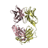
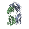
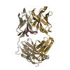
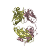
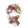
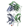
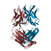
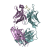
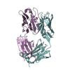
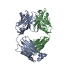
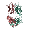
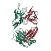
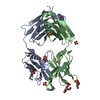
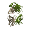
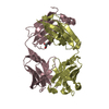
| Title | Crystal structure of the anti-estradiol antibody 57-2 | ||||||
 Components Components |
| ||||||
 Keywords Keywords |  IMMUNE SYSTEM / antibody / steroid IMMUNE SYSTEM / antibody / steroid | ||||||
| Function / homology |  Function and homology information Function and homology informationInitial triggering of complement / Classical antibody-mediated complement activation / FCGR activation / Role of phospholipids in phagocytosis / Regulation of Complement cascade / Regulation of actin dynamics for phagocytic cup formation / humoral immune response mediated by circulating immunoglobulin / phagocytosis, recognition / positive regulation of type IIa hypersensitivity / positive regulation of type I hypersensitivity ...Initial triggering of complement / Classical antibody-mediated complement activation / FCGR activation / Role of phospholipids in phagocytosis / Regulation of Complement cascade / Regulation of actin dynamics for phagocytic cup formation / humoral immune response mediated by circulating immunoglobulin / phagocytosis, recognition / positive regulation of type IIa hypersensitivity / positive regulation of type I hypersensitivity / antibody-dependent cellular cytotoxicity / Fc-gamma receptor I complex binding / phagocytosis, engulfment / IgG immunoglobulin complex / immunoglobulin mediated immune response / immunoglobulin complex, circulating / immunoglobulin receptor binding / positive regulation of phagocytosis / B cell differentiation / complement activation, classical pathway / antigen binding / positive regulation of immune response / antibacterial humoral response / blood microparticle / defense response to bacterium / immune response / external side of plasma membrane / extracellular space / extracellular exosome / metal ion binding / plasma membrane / cytoplasmSimilarity search - Function | ||||||
| Biological species |  Mus musculus (house mouse) Mus musculus (house mouse) | ||||||
| Method | X-RAY DIFFRACTION / MOLECULAR REPLACEMENT / Resolution: 2.51 Å | ||||||
 Authors Authors | Lamminmaki, U. / Kankare, J.A. | ||||||
 Citation Citation | Journal: J.Biol.Chem. / Year: 2001 Title: Crystal structure of a recombinant anti-estradiol Fab fragment in complex with 17beta -estradiol. Authors: Lamminmaki, U. / Kankare, J.A. #1: Journal: Acta Crystallogr.,Sect.D / Year: 2000Title: Crystallization and preliminary X-ray analysis of a recombinant Fab fragment in complex with 17-beta-estradiol Authors: Lamminmaki, U. / Kankare, J.A. | ||||||
| History |
|
- Structure visualization
| Structure viewer | Molecule: MolmilJmol/JSmol |
|---|
- Downloads & links
-Download
| PDBx/mmCIF format | 1jhk.cif.gz | 97 KB | Display | PDBx/mmCIF format |
|---|---|---|---|---|
| PDB format | pdb1jhk.ent.gz | 73 KB | Display | PDB format |
| PDBx/mmJSON format | 1jhk.json.gz | Tree view | PDBx/mmJSON format | |
| Others |  Other downloads Other downloads |
-Validation report
| Arichive directory | https://data.pdbj.org/pub/pdb/validation_reports/jh/1jhkftp://data.pdbj.org/pub/pdb/validation_reports/jh/1jhk | HTTPS FTP |
|---|
-Related structure data
| Related structure data | 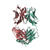 1jglC 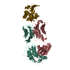 1fdlS 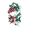 1tetS C: citing same article ( S: Starting model for refinement |
|---|---|
| Similar structure data |


-Links
 PDBj
PDBj


- Assembly
| Deposited unit | 
| ||||||||
|---|---|---|---|---|---|---|---|---|---|
| 1 |
| ||||||||
| Unit cell |
|
-Components
| #1: Antibody | Mass: 23665.074 Da / Num. of mol.: 1 Source method: isolated from a genetically manipulated source Source: (gene. exp.) Mus musculus (house mouse) / Plasmid: pCOMB3 / Production host:  Escherichia coli (E. coli) / Strain (production host): XL-1 blue / References: UniProt: Q8VCP0, UniProt: Q7TS98*PLUS Escherichia coli (E. coli) / Strain (production host): XL-1 blue / References: UniProt: Q8VCP0, UniProt: Q7TS98*PLUS |
|---|---|
| #2: Antibody | Mass: 23460.309 Da / Num. of mol.: 1 / Fragment: residues 1-215 Source method: isolated from a genetically manipulated source Source: (gene. exp.) Mus musculus (house mouse) / Plasmid: pCOMB3 / Production host: Escherichia coli (E. coli) / Strain (production host): XL-1 blue / References: UniProt: P01869, UniProt: Q99LC4*PLUS |
| #3: Water | ChemComp-HOH / Water Mass: 18.015 Da / Num. of mol.: 162 / Source method: isolated from a natural source / Formula: H2O Mass: 18.015 Da / Num. of mol.: 162 / Source method: isolated from a natural source / Formula: H2O |
-Experimental details
-Experiment
| Experiment | Method: X-RAY DIFFRACTION / Number of used crystals: 1 |
|---|
- Sample preparation
| Crystal | Density Matthews: 2.73 Å3/Da / Density % sol: 55 % | ||||||||||||||||||||||||
|---|---|---|---|---|---|---|---|---|---|---|---|---|---|---|---|---|---|---|---|---|---|---|---|---|---|
| Crystal grow | Temperature: 277 K / Method: vapor diffusion, sitting drop / pH: 9.1 Details: PEG4000, PEG8000, Tris-HCl, pH 9.1, VAPOR DIFFUSION, SITTING DROP, temperature 277K | ||||||||||||||||||||||||
| Crystal grow | *PLUS Details: used microseeding / PH range low: 9.5 / PH range high: 9 | ||||||||||||||||||||||||
| Components of the solutions | *PLUS
|
-Data collection
| Diffraction | Mean temperature: 100 K |
|---|---|
| Diffraction source | Source: ROTATING ANODE / Type: RIGAKU RU200HB / Wavelength: 1.5418 Å |
| Detector | Type: RIGAKU RAXIS IIC / Detector: IMAGE PLATE |
| Radiation | Monochromator: GRAPHITE / Protocol: SINGLE WAVELENGTH / Monochromatic (M) / Laue (L): M / Scattering type: x-ray |
| Radiation wavelength | Wavelength: 1.5418 Å / Relative weight: 1 |
| Reflection | Resolution: 2.5→50 Å / Num. all: 17081 / Num. obs: 17081 / % possible obs: 93.4 % / Observed criterion σ(F): 0 / Observed criterion σ(I): 0 / Redundancy: 4.3 % / Biso Wilson estimate: 36 Å2 / Rmerge(I) obs: 0.052 |
| Reflection shell | Resolution: 2.5→2.59 Å / Redundancy: 1.9 % / Rmerge(I) obs: 0.078 / % possible all: 72.7 |
| Reflection | *PLUS Lowest resolution: 50 Å / % possible obs: 91.8 % |
| Reflection shell | *PLUS % possible obs: 72.7 % |
- Processing
| Software |
| |||||||||||||||||||||||||
|---|---|---|---|---|---|---|---|---|---|---|---|---|---|---|---|---|---|---|---|---|---|---|---|---|---|---|
| Refinement | Method to determine structure: MOLECULAR REPLACEMENT Starting model: Combination of the PDB entries 1TET (variable heavy domain) and 1FDL (variable light domain and both constant domains) Resolution: 2.51→46.34 Å / Rfactor Rfree error: 0.008 / Data cutoff high absF: 650567.05 / Data cutoff low absF: 0 / Isotropic thermal model: RESTRAINED / Cross valid method: THROUGHOUT / σ(F): 0 / Stereochemistry target values: Engh & Huber
| |||||||||||||||||||||||||
| Solvent computation | Solvent model: FLAT MODEL / Bsol: 33.58 Å2 / ksol: 0.357 e/Å3 | |||||||||||||||||||||||||
| Displacement parameters | Biso mean: 33.2 Å2
| |||||||||||||||||||||||||
| Refine analyze |
| |||||||||||||||||||||||||
| Refinement step | Cycle: LAST / Resolution: 2.51→46.34 Å
| |||||||||||||||||||||||||
| Refine LS restraints |
| |||||||||||||||||||||||||
| LS refinement shell | Resolution: 2.5→2.66 Å / Rfactor Rfree error: 0.027 / Total num. of bins used: 6
| |||||||||||||||||||||||||
| Xplor file |
| |||||||||||||||||||||||||
| Software | *PLUS Name: CNS / Version: 1 / Classification: refinement | |||||||||||||||||||||||||
| Refinement | *PLUS σ(F): 0 / % reflection Rfree: 6.9 % / Rfactor all: 0.23 / Rfactor obs: 0.201 / Rfactor Rfree: 0.252 | |||||||||||||||||||||||||
| Solvent computation | *PLUS | |||||||||||||||||||||||||
| Displacement parameters | *PLUS Biso mean: 33.2 Å2 | |||||||||||||||||||||||||
| Refine LS restraints | *PLUS
| |||||||||||||||||||||||||
| LS refinement shell | *PLUS Rfactor Rfree: 0.344 / % reflection Rfree: 7 % / Rfactor Rwork: 0.248 / Rfactor obs: 0.258 |
