 Movie
Movie Controller

[English] 日本語
 Yorodumi
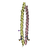
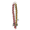
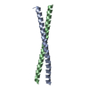
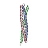
- PDB-1jcc: Crystal Structure of a Novel Alanine-Zipper Trimer at 1.7 A Resol... -
+ Open data

- Basic information
| Entry | Database: PDB / ID: 1jcc | ||||||
|---|---|---|---|---|---|---|---|
| Title | Crystal Structure of a Novel Alanine-Zipper Trimer at 1.7 A Resolution, V13A,L16A,V20A,L23A,V27A,M30A,V34A mutations | ||||||
 Components Components | MAJOR OUTER MEMBRANE LIPOPROTEIN | ||||||
 Keywords Keywords |  MEMBRANE PROTEIN / LIPOPROTEIN / PROTEIN FOLDING / COILED COIL / HELIX CAPPING / ALANINE-ZIPPER MEMBRANE PROTEIN / LIPOPROTEIN / PROTEIN FOLDING / COILED COIL / HELIX CAPPING / ALANINE-ZIPPER | ||||||
| Function / homology |  Function and homology information Function and homology informationperiplasmic space organization / lipid modification / peptidoglycan binding / cell outer membrane / outer membrane-bounded periplasmic space / lipid binding / extracellular region / membrane / identical protein bindingSimilarity search - Function | ||||||
| Biological species |  Escherichia coli (E. coli) Escherichia coli (E. coli) | ||||||
| Method | X-RAY DIFFRACTION / SYNCHROTRON / MOLECULAR REPLACEMENT / Resolution: 1.7 Å | ||||||
 Authors Authors | Liu, J. / Dai, J. / Lu, M. | ||||||
 Citation Citation | Journal: Biochemistry / Year: 2003 Title: Zinc-Mediated Helix Capping in A Triple-Helical Protein Authors: Liu, J. / Dai, J. / Lu, M. | ||||||
| History |
|
- Structure visualization
| Structure viewer | Molecule: MolmilJmol/JSmol |
|---|
- Downloads & links
-Download
| PDBx/mmCIF format | 1jcc.cif.gz | 45.3 KB | Display | PDBx/mmCIF format |
|---|---|---|---|---|
| PDB format | pdb1jcc.ent.gz | 31.8 KB | Display | PDB format |
| PDBx/mmJSON format | 1jcc.json.gz | Tree view | PDBx/mmJSON format | |
| Others |  Other downloads Other downloads |
-Validation report
| Arichive directory | https://data.pdbj.org/pub/pdb/validation_reports/jc/1jccftp://data.pdbj.org/pub/pdb/validation_reports/jc/1jcc | HTTPS FTP |
|---|
-Related structure data
| Related structure data | 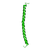 1eq7S  1jcb S: Starting model for refinement |
|---|---|
| Similar structure data |


-Links
 PDBj
PDBj


- Assembly
| Deposited unit | 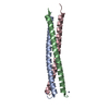
| ||||||||
|---|---|---|---|---|---|---|---|---|---|
| 1 |
| ||||||||
| Unit cell |
| ||||||||
| Details | The biological assembly is a trimer. |
-Components
| #1: Protein | Mass: 5905.234 Da / Num. of mol.: 3 / Mutation: V13A,L16A,V20A,L23A,V27A,M30A,V34A Source method: isolated from a genetically manipulated source Source: (gene. exp.) Escherichia coli (E. coli) / Production host: Escherichia coli (E. coli) / References: UniProt: P69776#2: Chemical |   Mass: 65.409 Da / Num. of mol.: 3 / Source method: obtained synthetically / Formula: Zn Mass: 65.409 Da / Num. of mol.: 3 / Source method: obtained synthetically / Formula: Zn#3: Water | ChemComp-HOH / | Water Mass: 18.015 Da / Num. of mol.: 234 / Source method: isolated from a natural source / Formula: H2O Mass: 18.015 Da / Num. of mol.: 234 / Source method: isolated from a natural source / Formula: H2O |
|---|
-Experimental details
-Experiment
| Experiment | Method: X-RAY DIFFRACTION / Number of used crystals: 1 |
|---|
- Sample preparation
| Crystal | Density Matthews: 2.06 Å3/Da / Density % sol: 40.33 % | ||||||||||||||||||||||||||||||||||||
|---|---|---|---|---|---|---|---|---|---|---|---|---|---|---|---|---|---|---|---|---|---|---|---|---|---|---|---|---|---|---|---|---|---|---|---|---|---|
| Crystal grow | Temperature: 293 K / Method: vapor diffusion, hanging drop / pH: 5.6 Details: Sodium acetate, zinc acetate, PEG 8000, pH 5.6, VAPOR DIFFUSION, HANGING DROP, temperature 293K | ||||||||||||||||||||||||||||||||||||
| Crystal grow | *PLUS Temperature: 4 ℃ / Method: vapor diffusion, hanging drop | ||||||||||||||||||||||||||||||||||||
| Components of the solutions | *PLUS
|
-Data collection
| Diffraction | Mean temperature: 95 K |
|---|---|
| Diffraction source | Source: SYNCHROTRON / Site: NSLS  / Beamline: X26C / Wavelength: 1.1 / Wavelength: 1.1 Å / Beamline: X26C / Wavelength: 1.1 / Wavelength: 1.1 Å |
| Detector | Type: ADSC QUANTUM 4 / Detector: CCD / Date: Apr 20, 2000 |
| Radiation | Monochromator: GRAPHITE / Protocol: SINGLE WAVELENGTH / Monochromatic (M) / Laue (L): M / Scattering type: x-ray |
| Radiation wavelength | Wavelength: 1.1 Å / Relative weight: 1 |
| Reflection | Resolution: 1.7→50 Å / Num. all: 15821 / Num. obs: 15821 / % possible obs: 97.6 % / Observed criterion σ(F): 0 / Observed criterion σ(I): 0 / Redundancy: 2.5 % / Biso Wilson estimate: 21.9 Å2 / Rmerge(I) obs: 0.079 / Net I/σ(I): 7.7 |
| Reflection shell | Resolution: 1.7→1.76 Å / Redundancy: 2.5 % / Rmerge(I) obs: 0.383 / Mean I/σ(I) obs: 3.3 / Num. unique all: 1605 / % possible all: 97.6 |
| Reflection | *PLUS Lowest resolution: 50 Å / Num. measured all: 69970 |
- Processing
| Software |
| ||||||||||||||||||||||||||||||||||||
|---|---|---|---|---|---|---|---|---|---|---|---|---|---|---|---|---|---|---|---|---|---|---|---|---|---|---|---|---|---|---|---|---|---|---|---|---|---|
| Refinement | Method to determine structure: MOLECULAR REPLACEMENT Starting model: PDB ENTRY 1EQ7 Resolution: 1.7→35.13 Å / Rfactor Rfree error: 0.006 / Data cutoff high absF: 1029074.03 / Data cutoff low absF: 0 / Isotropic thermal model: RESTRAINED / Cross valid method: THROUGHOUT / σ(F): 0 / σ(I): 0 / Stereochemistry target values: Engh & Huber
| ||||||||||||||||||||||||||||||||||||
| Solvent computation | Solvent model: FLAT MODEL / Bsol: 73.9 Å2 / ksol: 0.408 e/Å3 | ||||||||||||||||||||||||||||||||||||
| Displacement parameters | Biso mean: 22.1 Å2
| ||||||||||||||||||||||||||||||||||||
| Refine analyze |
| ||||||||||||||||||||||||||||||||||||
| Refinement step | Cycle: LAST / Resolution: 1.7→35.13 Å
| ||||||||||||||||||||||||||||||||||||
| Refine LS restraints |
| ||||||||||||||||||||||||||||||||||||
| LS refinement shell | Resolution: 1.7→1.81 Å / Rfactor Rfree error: 0.019 / Total num. of bins used: 6
| ||||||||||||||||||||||||||||||||||||
| Xplor file |
| ||||||||||||||||||||||||||||||||||||
| Refinement | *PLUS Highest resolution: 1.7 Å / Lowest resolution: 50 Å / Num. reflection obs: 14227 | ||||||||||||||||||||||||||||||||||||
| Solvent computation | *PLUS | ||||||||||||||||||||||||||||||||||||
| Displacement parameters | *PLUS | ||||||||||||||||||||||||||||||||||||
| Refine LS restraints | *PLUS
|
