 Movie
Movie Controller


+ Open data

- Basic information
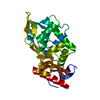
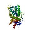
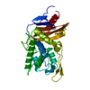
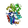
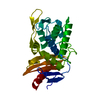
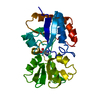
| Entry | Database: PDB / ID: 1j9m | |||||||||
|---|---|---|---|---|---|---|---|---|---|---|
| Title | K38H mutant of Streptomyces K15 DD-transpeptidase | |||||||||
 Components Components | DD-transpeptidase | |||||||||
 Keywords Keywords | HYDROLASE / penicillin-binding / DD-transpeptidase / serine peptidase / beta-lactamase / hydrolase carboxypeptidase | |||||||||
| Function / homology |  Function and homology informationserine-type D-Ala-D-Ala carboxypeptidase / serine-type D-Ala-D-Ala carboxypeptidase activity / peptidoglycan biosynthetic process / cell wall organization / regulation of cell shape / proteolysis / extracellular region Function and homology informationserine-type D-Ala-D-Ala carboxypeptidase / serine-type D-Ala-D-Ala carboxypeptidase activity / peptidoglycan biosynthetic process / cell wall organization / regulation of cell shape / proteolysis / extracellular regionSimilarity search - Function | |||||||||
| Biological species |  Streptomyces sp. (bacteria) Streptomyces sp. (bacteria) | |||||||||
| Method | X-RAY DIFFRACTION / SYNCHROTRON / MOLECULAR REPLACEMENT / Resolution: 1.65 Å | |||||||||
 Authors Authors | Fonze, E. / Rhazi, N. / Nguyen-Disteche, M. / Charlier, P. | |||||||||
 Citation Citation | Journal: Biochemistry / Year: 2003 Title: Catalytic mechanism of the Streptomyces K15 DD-transpeptidase/penicillin-binding protein probed by site-directed mutagenesis and structural analysis. Authors: Rhazi, N. / Charlier, P. / Dehareng, D. / Engher, D. / Vermeire, M. / Frere, J.M. / Nguyen-Disteche, M. / Fonze, E. #1: Journal: J.Biol.Chem. / Year: 1999Title: The Crystal Structure of a Penicilloyl-Serine Transferase of Intermediate Penicillin Sensitivity Authors: Fonze, E. / Vermeire, M. / Nguyen-Disteche, M. / Brasseur, R. / Charlier, P. #2: Journal: J.Mol.Biol. / Year: 1994Title: Crystallization and X-Ray Diffraction Study of the Streptomyces K15 Penicillin-Binding DD-Transpeptidase Authors: Englebert, S. / Charlier, P. / Fonze, E. / To'th, Y. / Vermeire, M. / Van Beeumen, J. / Grandchamps, J. / Hoffmann, K. / Leyh-Bouille, M. / Nguyen-Disteche, M. / Ghuysen, J.-M. | |||||||||
| History |
|
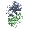
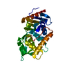
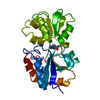
- Structure visualization
| Structure viewer | Molecule: MolmilJmol/JSmol |
|---|
- Downloads & links
-Download
| PDBx/mmCIF format | 1j9m.cif.gz | 64.3 KB | Display | PDBx/mmCIF format |
|---|---|---|---|---|
| PDB format | pdb1j9m.ent.gz | 46.3 KB | Display | PDB format |
| PDBx/mmJSON format | 1j9m.json.gz | Tree view | PDBx/mmJSON format | |
| Others |  Other downloads Other downloads |
-Validation report
| Arichive directory | https://data.pdbj.org/pub/pdb/validation_reports/j9/1j9mftp://data.pdbj.org/pub/pdb/validation_reports/j9/1j9m | HTTPS FTP |
|---|
-Related structure data


-Links
 PDBj
PDBj
- Assembly
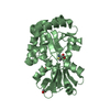
| Deposited unit | 
| ||||||||
|---|---|---|---|---|---|---|---|---|---|
| 1 |
| ||||||||
| Unit cell |
| ||||||||
| Details | The biological assembly is a monomer |
-Components
| #1: Protein | Mass: 27517.287 Da / Num. of mol.: 1 / Mutation: K38H Source method: isolated from a genetically manipulated source Source: (gene. exp.) Streptomyces sp. (bacteria) / Strain: K15 / Species (production host): Streptomyces lividans / Production host: Streptomyces lividans TK24 (bacteria) / Strain (production host): TK24References: UniProt: P39042, serine-type D-Ala-D-Ala carboxypeptidase |
|---|---|
| #2: Chemical | ChemComp-NA /   Mass: 22.990 Da / Num. of mol.: 1 / Source method: obtained synthetically / Formula: Na Mass: 22.990 Da / Num. of mol.: 1 / Source method: obtained synthetically / Formula: Na |
| #3: Chemical | ChemComp-CL / Chloride  Mass: 35.453 Da / Num. of mol.: 1 / Source method: obtained synthetically / Formula: Cl Mass: 35.453 Da / Num. of mol.: 1 / Source method: obtained synthetically / Formula: Cl |
| #4: Water | ChemComp-HOH / Water Mass: 18.015 Da / Num. of mol.: 197 / Source method: isolated from a natural source / Formula: H2O Mass: 18.015 Da / Num. of mol.: 197 / Source method: isolated from a natural source / Formula: H2O |
-Experimental details
-Experiment
| Experiment | Method: X-RAY DIFFRACTION / Number of used crystals: 1 |
|---|
- Sample preparation
| Crystal | Density Matthews: 2.33 Å3/Da / Density % sol: 47.12 % | ||||||||||||||||||||||||||||||
|---|---|---|---|---|---|---|---|---|---|---|---|---|---|---|---|---|---|---|---|---|---|---|---|---|---|---|---|---|---|---|---|
| Crystal grow | Temperature: 293 K / Method: vapor diffusion, hanging drop / pH: 7.2 Details: Tris 0.1 M, PEG 6K 30%, NaCl 0.4 M, pH 7.2, VAPOR DIFFUSION, HANGING DROP, temperature 293K | ||||||||||||||||||||||||||||||
| Crystal grow | *PLUS Temperature: 20 ℃ | ||||||||||||||||||||||||||||||
| Components of the solutions | *PLUS
|
-Data collection
| Diffraction | Mean temperature: 100 K |
|---|---|
| Diffraction source | Source: SYNCHROTRON / Site: ESRF  / Beamline: ID14-3 / Wavelength: 0.933 Å / Beamline: ID14-3 / Wavelength: 0.933 Å |
| Detector | Type: MARRESEARCH / Detector: CCD / Date: Jun 5, 1999 |
| Radiation | Protocol: SINGLE WAVELENGTH / Monochromatic (M) / Laue (L): M / Scattering type: x-ray |
| Radiation wavelength | Wavelength: 0.933 Å / Relative weight: 1 |
| Reflection | Resolution: 1.56→34.71 Å / Num. all: 37427 / Num. obs: 34332 / % possible obs: 85.9 % / Observed criterion σ(F): 0 / Observed criterion σ(I): 0 / Redundancy: 3.7 % / Biso Wilson estimate: 17.1 Å2 / Rmerge(I) obs: 0.038 / Net I/σ(I): 19.13 |
| Reflection shell | Resolution: 1.56→1.6 Å / Redundancy: 1.8 % / Rmerge(I) obs: 0.139 / % possible all: 39.7 |
| Reflection | *PLUS % possible obs: 91.7 % |
| Reflection shell | *PLUS % possible obs: 39.7 % / Num. unique obs: 1069 / Mean I/σ(I) obs: 4.1 |
- Processing
| Software |
| ||||||||||||||||||||||||||||||||||||||||
|---|---|---|---|---|---|---|---|---|---|---|---|---|---|---|---|---|---|---|---|---|---|---|---|---|---|---|---|---|---|---|---|---|---|---|---|---|---|---|---|---|---|
| Refinement | Method to determine structure: MOLECULAR REPLACEMENT / Resolution: 1.65→8 Å / Rfactor Rfree error: 0.007 / Data cutoff high absF: 100000 / Data cutoff low absF: 0.1 / Isotropic thermal model: RESTRAINED / σ(F): 2 Details: THE ELECTRON DENSITY FOR RESIDUES ILE 145, GLY 146 and ASN 147 IS NOT WELL-DEFINED. THOSE RESIDUES WERE EXCLUDED FROM THE REFINEMENT PROCESS.
| ||||||||||||||||||||||||||||||||||||||||
| Displacement parameters | Biso mean: 15 Å2
| ||||||||||||||||||||||||||||||||||||||||
| Refine analyze |
| ||||||||||||||||||||||||||||||||||||||||
| Refinement step | Cycle: LAST / Resolution: 1.65→8 Å
| ||||||||||||||||||||||||||||||||||||||||
| Refine LS restraints |
| ||||||||||||||||||||||||||||||||||||||||
| LS refinement shell | Resolution: 1.65→1.71 Å / Rfactor Rfree error: 0.025 / Total num. of bins used: 10
| ||||||||||||||||||||||||||||||||||||||||
| Software | *PLUS Name: X-PLOR(ONLINE) / Version: 3.851 / Classification: refinement | ||||||||||||||||||||||||||||||||||||||||
| Refinement | *PLUS σ(F): 2 / % reflection Rfree: 5 % | ||||||||||||||||||||||||||||||||||||||||
| Solvent computation | *PLUS | ||||||||||||||||||||||||||||||||||||||||
| Displacement parameters | *PLUS Biso mean: 15 Å2 | ||||||||||||||||||||||||||||||||||||||||
| Refine LS restraints | *PLUS
| ||||||||||||||||||||||||||||||||||||||||
| LS refinement shell | *PLUS Rfactor Rfree: 0.285 / % reflection Rfree: 4.8 % / Rfactor Rwork: 0.334 |
