 Movie
Movie Controller

[English] 日本語
 Yorodumi
- PDB-1j72: Crystal Structure of Mutant Macrophage Capping Protein (Cap G) wi... -
+ Open data

- Basic information
| Entry | Database: PDB / ID: 1j72 | ||||||
|---|---|---|---|---|---|---|---|
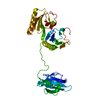
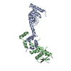
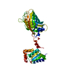
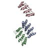
| Title | Crystal Structure of Mutant Macrophage Capping Protein (Cap G) with Actin-severing Activity in the Ca2+-Free Form | ||||||
 Components Components | Macrophage capping protein | ||||||
 Keywords Keywords |  STRUCTURAL PROTEIN / actin / human / capping / cap g / macrophage / gCap39 / Mbhl / gelsolin STRUCTURAL PROTEIN / actin / human / capping / cap g / macrophage / gCap39 / Mbhl / gelsolin | ||||||
| Function / homology |  Function and homology information Function and homology informationF-actin capping protein complex / actin filament severing / barbed-end actin filament capping / actin polymerization or depolymerization / cell projection assembly / Flemming body / ruffle / phosphatidylinositol-4,5-bisphosphate binding / centriole / central nervous system development ...F-actin capping protein complex / actin filament severing / barbed-end actin filament capping / actin polymerization or depolymerization / cell projection assembly / Flemming body / ruffle / phosphatidylinositol-4,5-bisphosphate binding / centriole / central nervous system development / mitotic spindle / actin filament binding / melanosome / actin cytoskeleton / lamellipodium / protein-containing complex assembly / cadherin binding / protein domain specific binding / protein-containing complex binding / nucleolus / extracellular exosome / nucleoplasm / nucleus / cytoplasmSimilarity search - Function | ||||||
| Biological species |  Homo sapiens (human) Homo sapiens (human) | ||||||
| Method | X-RAY DIFFRACTION / SYNCHROTRON / MIR / Resolution: 2.5 Å | ||||||
 Authors Authors | Vorobiev, S.M. / Southwick, F.S. / Storokopytov, B. / Almo, S.C. | ||||||
 Citation Citation | Journal: To be Published Title: Crystal Structure of Mutant Human Macrophage Capping Protein Authors: Vorobiev, S.M. / Southwick, F.S. / Storokopytov, B. / Almo, S.C. #1: Journal: J.Biol.Chem. / Year: 1995Title: Gain-of-function mutations conferring actin-severing activity to human macrophage Cap G Authors: Southwick, F.S. | ||||||
| History |
|
- Structure visualization
| Structure viewer | Molecule: MolmilJmol/JSmol |
|---|
- Downloads & links
-Download
| PDBx/mmCIF format | 1j72.cif.gz | 78.1 KB | Display | PDBx/mmCIF format |
|---|---|---|---|---|
| PDB format | pdb1j72.ent.gz | 58.6 KB | Display | PDB format |
| PDBx/mmJSON format | 1j72.json.gz | Tree view | PDBx/mmJSON format | |
| Others |  Other downloads Other downloads |
-Validation report
| Arichive directory | https://data.pdbj.org/pub/pdb/validation_reports/j7/1j72ftp://data.pdbj.org/pub/pdb/validation_reports/j7/1j72 | HTTPS FTP |
|---|
-Related structure data


-Links
 PDBj
PDBj
- Assembly
| Deposited unit | 
| ||||||||
|---|---|---|---|---|---|---|---|---|---|
| 1 |
| ||||||||
| 2 | 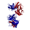
| ||||||||
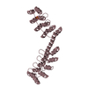
| Unit cell |
|
-Components
| #1: Protein | Mass: 38625.477 Da / Num. of mol.: 1 Mutation: 108HLNTLLGE -> 108QLDDYLGG; 148AFHTSTGAPAAIKK -> 148GFKHVVPNEVVVQR Source method: isolated from a genetically manipulated source Source: (gene. exp.) Homo sapiens (human) / Species (production host): Escherichia coli / Production host:  Escherichia coli BL21(DE3) (bacteria) / Strain (production host): BL21(DE3) / References: UniProt: P40121 Escherichia coli BL21(DE3) (bacteria) / Strain (production host): BL21(DE3) / References: UniProt: P40121 |
|---|---|
| #2: Water | ChemComp-HOH / Water Mass: 18.015 Da / Num. of mol.: 82 / Source method: isolated from a natural source / Formula: H2O Mass: 18.015 Da / Num. of mol.: 82 / Source method: isolated from a natural source / Formula: H2O |
-Experimental details
-Experiment
| Experiment | Method: X-RAY DIFFRACTION / Number of used crystals: 1 |
|---|
- Sample preparation
| Crystal | Density Matthews: 4.48 Å3/Da / Density % sol: 72.54 % |
|---|---|
| Crystal grow | Temperature: 298 K / Method: vapor diffusion, hanging drop / pH: 7 Details: 0.1M Tris-HCl, pH 7.0-8.0, 3.5 - 4.0 ammonium formate, VAPOR DIFFUSION, HANGING DROP, temperature 298K |
-Data collection
| Diffraction | Mean temperature: 100 K |
|---|---|
| Diffraction source | Source: SYNCHROTRON / Site: NSLS  / Beamline: X9B / Wavelength: 0.97178 Å / Beamline: X9B / Wavelength: 0.97178 Å |
| Detector | Type: ADSC QUANTUM 4 / Detector: CCD / Date: Jan 20, 1999 |
| Radiation | Protocol: SINGLE WAVELENGTH / Monochromatic (M) / Laue (L): M / Scattering type: x-ray |
| Radiation wavelength | Wavelength: 0.97178 Å / Relative weight: 1 |
| Reflection | Resolution: 2.5→15 Å / Num. all: 24873 / Num. obs: 24873 / % possible obs: 98.5 % / Observed criterion σ(F): 0 / Observed criterion σ(I): 0 / Redundancy: 5.86 % / Biso Wilson estimate: 36.5 Å2 / Rmerge(I) obs: 0.097 |
| Reflection shell | Resolution: 2.5→2.53 Å / Redundancy: 3.54 % / Rmerge(I) obs: 0.431 / Num. unique all: 1051 / % possible all: 85.5 |
- Processing
| Software |
| ||||||||||||||||||||||||||||||||||||||||||||||||||||||||||||||||||||||||||||||||
|---|---|---|---|---|---|---|---|---|---|---|---|---|---|---|---|---|---|---|---|---|---|---|---|---|---|---|---|---|---|---|---|---|---|---|---|---|---|---|---|---|---|---|---|---|---|---|---|---|---|---|---|---|---|---|---|---|---|---|---|---|---|---|---|---|---|---|---|---|---|---|---|---|---|---|---|---|---|---|---|---|---|
| Refinement | Method to determine structure: MIR / Resolution: 2.5→14.96 Å / Rfactor Rfree error: 0.01 / Data cutoff high absF: 2189205.08 / Data cutoff low absF: 0 / Isotropic thermal model: RESTRAINED / Cross valid method: THROUGHOUT / σ(F): 2
| ||||||||||||||||||||||||||||||||||||||||||||||||||||||||||||||||||||||||||||||||
| Solvent computation | Solvent model: FLAT MODEL / Bsol: 53.11 Å2 / ksol: 0.308 e/Å3 | ||||||||||||||||||||||||||||||||||||||||||||||||||||||||||||||||||||||||||||||||
| Displacement parameters | Biso mean: 65.8 Å2
| ||||||||||||||||||||||||||||||||||||||||||||||||||||||||||||||||||||||||||||||||
| Refine analyze |
| ||||||||||||||||||||||||||||||||||||||||||||||||||||||||||||||||||||||||||||||||
| Refinement step | Cycle: LAST / Resolution: 2.5→14.96 Å
| ||||||||||||||||||||||||||||||||||||||||||||||||||||||||||||||||||||||||||||||||
| Refine LS restraints |
| ||||||||||||||||||||||||||||||||||||||||||||||||||||||||||||||||||||||||||||||||
| LS refinement shell | Resolution: 2.5→2.66 Å / Rfactor Rfree error: 0.039 / Total num. of bins used: 6
|
