 Movie
Movie Controller

[English] 日本語
 Yorodumi
- PDB-1j4x: HUMAN VH1-RELATED DUAL-SPECIFICITY PHOSPHATASE C124S MUTANT-PEPTI... -
+ Open data

- Basic information
| Entry | Database: PDB / ID: 1j4x | |||||||||
|---|---|---|---|---|---|---|---|---|---|---|
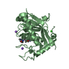
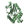
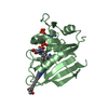
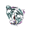
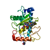
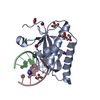
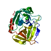
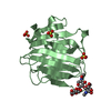
| Title | HUMAN VH1-RELATED DUAL-SPECIFICITY PHOSPHATASE C124S MUTANT-PEPTIDE COMPLEX | |||||||||
 Components Components |
| |||||||||
 Keywords Keywords |  HYDROLASE / PROTEIN DUAL-SPECIFICITY PHOSPHATASE HYDROLASE / PROTEIN DUAL-SPECIFICITY PHOSPHATASE | |||||||||
| Function / homology |  Function and homology informationMAP kinase phosphatase activity / negative regulation of chemotaxis / negative regulation of T cell activation / protein tyrosine/serine/threonine phosphatase activity / positive regulation of focal adhesion disassembly / negative regulation of JNK cascade / ERKs are inactivated / regulation of focal adhesion assembly / negative regulation of T cell receptor signaling pathway / negative regulation of MAPK cascade ...MAP kinase phosphatase activity / negative regulation of chemotaxis / negative regulation of T cell activation / protein tyrosine/serine/threonine phosphatase activity / positive regulation of focal adhesion disassembly / negative regulation of JNK cascade / ERKs are inactivated / regulation of focal adhesion assembly / negative regulation of T cell receptor signaling pathway / negative regulation of MAPK cascade / myosin phosphatase activity / peptidyl-tyrosine dephosphorylation involved in inactivation of protein kinase activity / protein-serine/threonine phosphatase / negative regulation of epidermal growth factor receptor signaling pathway / phosphatase activity / immunological synapse / peptidyl-tyrosine dephosphorylation / dephosphorylation / cellular response to epidermal growth factor stimulus / cytoskeletal protein binding / positive regulation of mitotic cell cycle / negative regulation of cell migration / protein tyrosine kinase binding / protein-tyrosine-phosphatase / protein tyrosine phosphatase activity / negative regulation of ERK1 and ERK2 cascade / receptor tyrosine kinase binding / protein kinase binding / nucleoplasm / nucleus / cytosol / cytoplasm Function and homology informationMAP kinase phosphatase activity / negative regulation of chemotaxis / negative regulation of T cell activation / protein tyrosine/serine/threonine phosphatase activity / positive regulation of focal adhesion disassembly / negative regulation of JNK cascade / ERKs are inactivated / regulation of focal adhesion assembly / negative regulation of T cell receptor signaling pathway / negative regulation of MAPK cascade ...MAP kinase phosphatase activity / negative regulation of chemotaxis / negative regulation of T cell activation / protein tyrosine/serine/threonine phosphatase activity / positive regulation of focal adhesion disassembly / negative regulation of JNK cascade / ERKs are inactivated / regulation of focal adhesion assembly / negative regulation of T cell receptor signaling pathway / negative regulation of MAPK cascade / myosin phosphatase activity / peptidyl-tyrosine dephosphorylation involved in inactivation of protein kinase activity / protein-serine/threonine phosphatase / negative regulation of epidermal growth factor receptor signaling pathway / phosphatase activity / immunological synapse / peptidyl-tyrosine dephosphorylation / dephosphorylation / cellular response to epidermal growth factor stimulus / cytoskeletal protein binding / positive regulation of mitotic cell cycle / negative regulation of cell migration / protein tyrosine kinase binding / protein-tyrosine-phosphatase / protein tyrosine phosphatase activity / negative regulation of ERK1 and ERK2 cascade / receptor tyrosine kinase binding / protein kinase binding / nucleoplasm / nucleus / cytosol / cytoplasmSimilarity search - Function | |||||||||
| Biological species |  Homo sapiens (human) Homo sapiens (human) | |||||||||
| Method | X-RAY DIFFRACTION / SYNCHROTRON / molecular replacement / Resolution: 2.75 Å | |||||||||
 Authors Authors | Schumacher, M.A. / Todd, J.L. / Tanner, K.G. / Denu, J.M. | |||||||||
 Citation Citation | Journal: Biochemistry / Year: 2002 Title: Structural basis for the recognition of a bisphosphorylated MAP kinase peptide by human VHR protein Phosphatase. Authors: Schumacher, M.A. / Todd, J.L. / Rice, A.E. / Tanner, K.G. / Denu, J.M. | |||||||||
| History |
|
- Structure visualization
| Structure viewer | Molecule: MolmilJmol/JSmol |
|---|
- Downloads & links
-Download
| PDBx/mmCIF format | 1j4x.cif.gz | 52 KB | Display | PDBx/mmCIF format |
|---|---|---|---|---|
| PDB format | pdb1j4x.ent.gz | 36.4 KB | Display | PDB format |
| PDBx/mmJSON format | 1j4x.json.gz | Tree view | PDBx/mmJSON format | |
| Others |  Other downloads Other downloads |
-Validation report
| Arichive directory | https://data.pdbj.org/pub/pdb/validation_reports/j4/1j4xftp://data.pdbj.org/pub/pdb/validation_reports/j4/1j4x | HTTPS FTP |
|---|
-Related structure data
| Similar structure data |
|---|


-Links
 PDBj
PDBj


- Assembly
| Deposited unit | 
| ||||||||
|---|---|---|---|---|---|---|---|---|---|
| 1 |
| ||||||||
| Unit cell |
|
-Components
| #1: Protein | Mass: 20354.012 Da / Num. of mol.: 1 / Mutation: YES Source method: isolated from a genetically manipulated source Source: (gene. exp.) Homo sapiens (human) / Plasmid: PT7-7-VHR / Species (production host): Escherichia coli / Production host:  Escherichia coli BL21(DE3) (bacteria) / Strain (production host): BL21(DE3) / References: UniProt: P51452, protein-tyrosine-phosphatase Escherichia coli BL21(DE3) (bacteria) / Strain (production host): BL21(DE3) / References: UniProt: P51452, protein-tyrosine-phosphatase |
|---|---|
| #2: Protein/peptide | Mass: 1414.283 Da / Num. of mol.: 1 / Source method: obtained synthetically |
| #3: Water | ChemComp-HOH / Water Mass: 18.015 Da / Num. of mol.: 66 / Source method: isolated from a natural source / Formula: H2O Mass: 18.015 Da / Num. of mol.: 66 / Source method: isolated from a natural source / Formula: H2O |
-Experimental details
-Experiment
| Experiment | Method: X-RAY DIFFRACTION / Number of used crystals: 1 |
|---|
- Sample preparation
| Crystal | Density Matthews: 2.23 Å3/Da / Density % sol: 44.76 % | ||||||||||||||||||||||||||||||
|---|---|---|---|---|---|---|---|---|---|---|---|---|---|---|---|---|---|---|---|---|---|---|---|---|---|---|---|---|---|---|---|
| Crystal grow | Temperature: 298 K / Method: vapor diffusion, hanging drop / pH: 7 Details: PEG 4000, ISOPROPANOL, pH 7.00, VAPOR DIFFUSION, HANGING DROP, temperature 298K | ||||||||||||||||||||||||||||||
| Crystal grow | *PLUS pH: 5.6 / Method: vapor diffusion | ||||||||||||||||||||||||||||||
| Components of the solutions | *PLUS
|
-Data collection
| Diffraction | Mean temperature: 298 K |
|---|---|
| Diffraction source | Source: SYNCHROTRON / Site: SSRL  / Beamline: BL7-1 / Wavelength: 1.08 / Beamline: BL7-1 / Wavelength: 1.08 |
| Detector | Type: MARRESEARCH / Detector: IMAGE PLATE / Date: Feb 20, 1997 |
| Radiation | Protocol: SINGLE WAVELENGTH / Monochromatic (M) / Laue (L): M / Scattering type: x-ray |
| Radiation wavelength | Wavelength: 1.08 Å / Relative weight: 1 |
| Reflection | Resolution: 2.75→30 Å / Num. obs: 9234 / % possible obs: 90 % / Observed criterion σ(I): 0 / Redundancy: 1.5 % / Biso Wilson estimate: 45 Å2 / Rmerge(I) obs: 0.095 / Net I/σ(I): 17.1 |
| Reflection shell | Resolution: 2.75→2.9 Å / Redundancy: 1.2 % / Rmerge(I) obs: 0.29 / % possible all: 80 |
| Reflection | *PLUS Highest resolution: 2.75 Å / Lowest resolution: 10 Å / Num. measured all: 16753 |
- Processing
| Software |
| ||||||||||||||||||||||||||||||
|---|---|---|---|---|---|---|---|---|---|---|---|---|---|---|---|---|---|---|---|---|---|---|---|---|---|---|---|---|---|---|---|
| Refinement | Method to determine structure: molecular replacement / Resolution: 2.75→10 Å / σ(F): 0 / Stereochemistry target values: ENGH & HUBER
| ||||||||||||||||||||||||||||||
| Refinement step | Cycle: LAST / Resolution: 2.75→10 Å
| ||||||||||||||||||||||||||||||
| Refine LS restraints |
| ||||||||||||||||||||||||||||||
| Software | *PLUS Name: TNT / Classification: refinement | ||||||||||||||||||||||||||||||
| Refinement | *PLUS Lowest resolution: 10 Å / % reflection Rfree: 10 % / Rfactor all: 0.192 / Rfactor obs: 0.188 / Rfactor Rfree: 0.264 | ||||||||||||||||||||||||||||||
| Solvent computation | *PLUS | ||||||||||||||||||||||||||||||
| Displacement parameters | *PLUS |
