 Movie
Movie Controller

[English] 日本語
 Yorodumi
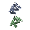
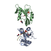
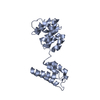
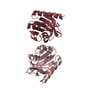
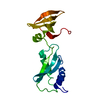
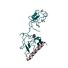
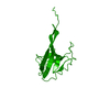
- PDB-1iuf: LOW RESOLUTION SOLUTION STRUCTURE OF THE TWO DNA-BINDING DOMAINS ... -
+ Open data

- Basic information
| Entry | Database: PDB / ID: 1iuf | ||||||
|---|---|---|---|---|---|---|---|
| Title | LOW RESOLUTION SOLUTION STRUCTURE OF THE TWO DNA-BINDING DOMAINS IN Schizosaccharomyces pombe ABP1 PROTEIN | ||||||
 Components Components | centromere abp1 protein | ||||||
 Keywords Keywords |  DNA BINDING PROTEIN / centromere / RIKEN Structural Genomics/Proteomics Initiative / RSGI / Structural Genomics DNA BINDING PROTEIN / centromere / RIKEN Structural Genomics/Proteomics Initiative / RSGI / Structural Genomics | ||||||
| Function / homology |  Function and homology information Function and homology informationprotein localization to mating-type region heterochromatin / donor selection / condensed chromosome, centromeric region => GO:0000779 / mating-type region heterochromatin / centromeric DNA binding / chromatin => GO:0000785 / mitotic sister chromatid segregation / sequence-specific DNA binding / DNA binding / nucleusSimilarity search - Function | ||||||
| Biological species |  Schizosaccharomyces pombe (fission yeast) Schizosaccharomyces pombe (fission yeast) | ||||||
| Method | SOLUTION NMR / torsion angle dynamics followed with simulated annealing | ||||||
 Authors Authors | Kikuchi, J. / Iwahara, J. / Kigawa, T. / Murakami, Y. / Okazaki, T. / Yokoyama, S. / RIKEN Structural Genomics/Proteomics Initiative (RSGI) | ||||||
 Citation Citation | Journal: J.Biomol.NMR / Year: 2002 Title: Solution structure determination of the two DNA-binding domains in the Schizosaccharomyces pombe Abp1 protein by a combination of dipolar coupling and diffusion anisotropy restraints. Authors: Kikuchi, J. / Iwahara, J. / Kigawa, T. / Murakami, Y. / Okazaki, T. / Yokoyama, S. | ||||||
| History |
|
- Structure visualization
| Structure viewer | Molecule: MolmilJmol/JSmol |
|---|
- Downloads & links
-Download
| PDBx/mmCIF format | 1iuf.cif.gz | 58.9 KB | Display | PDBx/mmCIF format |
|---|---|---|---|---|
| PDB format | pdb1iuf.ent.gz | 43.4 KB | Display | PDB format |
| PDBx/mmJSON format | 1iuf.json.gz | Tree view | PDBx/mmJSON format | |
| Others |  Other downloads Other downloads |
-Validation report
| Arichive directory | https://data.pdbj.org/pub/pdb/validation_reports/iu/1iufftp://data.pdbj.org/pub/pdb/validation_reports/iu/1iuf | HTTPS FTP |
|---|
-Related structure data
| Similar structure data | |
|---|---|
| Other databases |
-Links
 PDBj
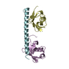
PDBj- Assembly
| Deposited unit | 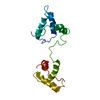
| |||||||||
|---|---|---|---|---|---|---|---|---|---|---|
| 1 |
| |||||||||
| NMR ensembles |
|
-Components
| #1: Protein | Mass: 17086.396 Da / Num. of mol.: 1 / Fragment: N-terminal domain Source method: isolated from a genetically manipulated source Source: (gene. exp.) Schizosaccharomyces pombe (fission yeast)Plasmid: pGADe / Species (production host): Escherichia coli / Production host:  Escherichia coli BL21 (bacteria) / Strain (production host): BL21 / References: UniProt: P49777 Escherichia coli BL21 (bacteria) / Strain (production host): BL21 / References: UniProt: P49777 |
|---|
-Experimental details
-Experiment
| Experiment | Method: SOLUTION NMR |
|---|---|
| NMR experiment | Type: 4D 13C/15N-separated NOESY |
| NMR details | Text: The structure was determined using triple-resonance NMR spectroscopy. |
- Sample preparation
| Details | Contents: 1mM Abp1 U-15N, 13C; 50mM Tris buffer; 90% H2O, 10% D2O |
|---|---|
| Sample conditions | Ionic strength: 50mM / pH: 6.8 / Pressure: 1 atm / Temperature: 310 K |
| Crystal grow | *PLUS Method: other / Details: NMR |
-NMR measurement
| Radiation | Protocol: SINGLE WAVELENGTH / Monochromatic (M) / Laue (L): M |
|---|---|
| Radiation wavelength | Relative weight: 1 |
| NMR spectrometer | Type: Bruker DRX / Manufacturer: Bruker / Model: DRX / Field strength: 600 MHz |
- Processing
| NMR software | Name: CNS / Version: 1 / Developer: Brunger, A.T. et al. / Classification: refinement |
|---|---|
| Refinement | Method: torsion angle dynamics followed with simulated annealing Software ordinal: 1 Details: residual sipolar coupling data were measured in pf1 phage liquid crystal diffusion anisotropy restraints were measured in solution |
| NMR ensemble | Conformer selection criteria: structures with the lowest energy Conformers calculated total number: 10 / Conformers submitted total number: 1 |
