 Movie
Movie Controller

[English] 日本語
 Yorodumi
- PDB-1ik6: 3D structure of the E1beta subunit of pyruvate dehydrogenase from... -
+ Open data

- Basic information
| Entry | Database: PDB / ID: 1ik6 | ||||||
|---|---|---|---|---|---|---|---|
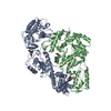
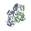
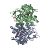
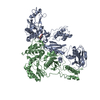
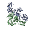
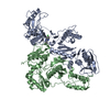
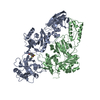
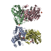
| Title | 3D structure of the E1beta subunit of pyruvate dehydrogenase from the archeon Pyrobaculum aerophilum | ||||||
 Components Components | pyruvate dehydrogenase | ||||||
 Keywords Keywords | OXIDOREDUCTASE / e1beta / pyruvate dehydrogenase / tetramer / gxxxg | ||||||
| Function / homology |  Function and homology information Function and homology information3-methyl-2-oxobutanoate dehydrogenase (2-methylpropanoyl-transferring) activity => GO:0003863 / 2-oxoacid oxidoreductase (ferredoxin) / branched-chain amino acid catabolic process / response to nutrient Similarity search - Function | ||||||
| Biological species |   Pyrobaculum aerophilum (archaea) Pyrobaculum aerophilum (archaea) | ||||||
| Method | X-RAY DIFFRACTION / SYNCHROTRON / MOLECULAR REPLACEMENT / Resolution: 2 Å | ||||||
 Authors Authors | Kleiger, G. / Perry, J. / Eisenberg, D. | ||||||
 Citation Citation | Journal: Biochemistry / Year: 2001 Title: 3D structure and significance of the GPhiXXG helix packing motif in tetramers of the E1beta subunit of pyruvate dehydrogenase from the archeon Pyrobaculum aerophilum. Authors: Kleiger, G. / Perry, J. / Eisenberg, D. | ||||||
| History |
| ||||||
| Remark 999 | SEQUENCE There is an N-terminal his tag that was NOT cleaved. However, it is not ordered so the ...SEQUENCE There is an N-terminal his tag that was NOT cleaved. However, it is not ordered so the author was not able to model it. |
- Structure visualization
| Structure viewer | Molecule: MolmilJmol/JSmol |
|---|
- Downloads & links
-Download
| PDBx/mmCIF format | 1ik6.cif.gz | 69.2 KB | Display | PDBx/mmCIF format |
|---|---|---|---|---|
| PDB format | pdb1ik6.ent.gz | 49.6 KB | Display | PDB format |
| PDBx/mmJSON format | 1ik6.json.gz | Tree view | PDBx/mmJSON format | |
| Others |  Other downloads Other downloads |
-Validation report
| Arichive directory | https://data.pdbj.org/pub/pdb/validation_reports/ik/1ik6ftp://data.pdbj.org/pub/pdb/validation_reports/ik/1ik6 | HTTPS FTP |
|---|
-Related structure data
| Related structure data | 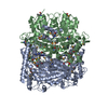 1qs0S S: Starting model for refinement |
|---|---|
| Similar structure data |
-Links
 PDBj
PDBj


- Assembly
| Deposited unit | 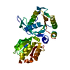
| ||||||||
|---|---|---|---|---|---|---|---|---|---|
| 1 | 
| ||||||||
| Unit cell |
| ||||||||
| Details | The biological assembly is a tetramer generated from the monomer in the asymmetric unit by the operations: -x,-y,z followed by -x,y,-z |
-Components
| #1: Protein | Mass: 39932.840 Da / Num. of mol.: 1 / Fragment: E1beta subunit Source method: isolated from a genetically manipulated source Source: (gene. exp.) Pyrobaculum aerophilum (archaea) / Plasmid: pET30Ek/LIC / Species (production host): Escherichia coli / Production host:  Escherichia coli BL21(DE3) (bacteria) / Strain (production host): BL21(DE3) / References: UniProt: Q8ZUR7 Escherichia coli BL21(DE3) (bacteria) / Strain (production host): BL21(DE3) / References: UniProt: Q8ZUR7 |
|---|---|
| #2: Water | ChemComp-HOH / Water Mass: 18.015 Da / Num. of mol.: 134 / Source method: isolated from a natural source / Formula: H2O Mass: 18.015 Da / Num. of mol.: 134 / Source method: isolated from a natural source / Formula: H2O |
-Experimental details
-Experiment
| Experiment | Method: X-RAY DIFFRACTION / Number of used crystals: 1 |
|---|
- Sample preparation
| Crystal | Density Matthews: 2.27 Å3/Da / Density % sol: 45.74 % | |||||||||||||||||||||||||||||||||||
|---|---|---|---|---|---|---|---|---|---|---|---|---|---|---|---|---|---|---|---|---|---|---|---|---|---|---|---|---|---|---|---|---|---|---|---|---|
| Crystal grow | Temperature: 291 K / Method: vapor diffusion, hanging drop / pH: 7.5 Details: Peg 400, Calcium Chloride, Sodium Hepes, pH 7.5, VAPOR DIFFUSION, HANGING DROP, temperature 291K | |||||||||||||||||||||||||||||||||||
| Crystal grow | *PLUS Temperature: 18 ℃ | |||||||||||||||||||||||||||||||||||
| Components of the solutions | *PLUS
|
-Data collection
| Diffraction | Mean temperature: 95 K |
|---|---|
| Diffraction source | Source: SYNCHROTRON / Site: NSLS  / Beamline: X8C / Wavelength: 1.072 Å / Beamline: X8C / Wavelength: 1.072 Å |
| Detector | Type: ADSC QUANTUM 4 / Detector: CCD / Date: Oct 14, 1999 |
| Radiation | Protocol: SINGLE WAVELENGTH / Monochromatic (M) / Laue (L): M / Scattering type: x-ray |
| Radiation wavelength | Wavelength: 1.072 Å / Relative weight: 1 |
| Reflection | Resolution: 2→50 Å / Num. all: 24820 / Num. obs: 22189 / % possible obs: 89.4 % / Observed criterion σ(F): 2 / Observed criterion σ(I): 2 / Redundancy: 5.6 % / Biso Wilson estimate: 25 Å2 / Rmerge(I) obs: 0.082 / Net I/σ(I): 22 |
| Reflection shell | Resolution: 2→2.07 Å / Redundancy: 5.6 % / Rmerge(I) obs: 0.257 / % possible all: 96 |
| Reflection | *PLUS Highest resolution: 2 Å / Lowest resolution: 50 Å |
| Reflection shell | *PLUS % possible obs: 96 % |
- Processing
| Software |
| ||||||||||||||||||||
|---|---|---|---|---|---|---|---|---|---|---|---|---|---|---|---|---|---|---|---|---|---|
| Refinement | Method to determine structure: MOLECULAR REPLACEMENT Starting model: PDB 1QS0 Beta-chain Resolution: 2→50 Å / σ(F): 0 / σ(I): 0 / Stereochemistry target values: Engh and Huber
| ||||||||||||||||||||
| Refinement step | Cycle: LAST / Resolution: 2→50 Å
| ||||||||||||||||||||
| Refine LS restraints |
| ||||||||||||||||||||
| LS refinement shell | Resolution: 2→2.02 Å /
| ||||||||||||||||||||
| Software | *PLUS Name: CNS / Classification: refinement | ||||||||||||||||||||
| Refinement | *PLUS Highest resolution: 2 Å / Lowest resolution: 50 Å / σ(F): 0 / Rfactor obs: 0.212 | ||||||||||||||||||||
| Solvent computation | *PLUS | ||||||||||||||||||||
| Displacement parameters | *PLUS | ||||||||||||||||||||
| Refine LS restraints | *PLUS Type: c_angle_deg / Dev ideal: 1.6 | ||||||||||||||||||||
| LS refinement shell | *PLUS Highest resolution: 2 Å / Rfactor Rfree: 0.321 / Rfactor Rwork: 0.251 |
