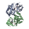
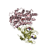
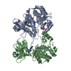
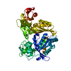
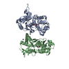
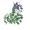
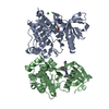
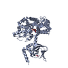
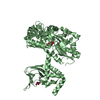
[English] 日本語
Yorodumi
- PDB-5jon: Crystal structure of the unliganded form of HCN2 CNBD -

+
Open data


ID or keywords:

Loading...

-
Basic information

Entry
Database: PDB / ID: 5jon
TitleCrystal structure of the unliganded form of HCN2 CNBD
ComponentsMaltose-binding periplasmic protein,Potassium/sodium hyperpolarization-activated cyclic nucleotide-gated channel 2
KeywordsTRANSPORT PROTEIN / HCN channels / cyclic nucleotide regulated channels / cyclic nucleotide binding domain
Function / homology
Function and homology information


HCN channels / HCN channel complex / intracellularly cAMP-activated cation channel activity / cellular response to cGMP / sodium ion import across plasma membrane / voltage-gated sodium channel activity / regulation of membrane depolarization / potassium ion import across plasma membrane / voltage-gated potassium channel activity / maltose binding ...HCN channels / HCN channel complex / intracellularly cAMP-activated cation channel activity / cellular response to cGMP / sodium ion import across plasma membrane / voltage-gated sodium channel activity / regulation of membrane depolarization / potassium ion import across plasma membrane / voltage-gated potassium channel activity / maltose binding / maltose transport / maltodextrin transmembrane transport / carbohydrate transmembrane transporter activity / sodium ion transmembrane transport / ATP-binding cassette (ABC) transporter complex, substrate-binding subunit-containing / cAMP binding / cellular response to cAMP / potassium ion transmembrane transport / somatodendritic compartment / dendrite membrane / dendritic shaft / regulation of membrane potential / PDZ domain binding / outer membrane-bounded periplasmic space / molecular adaptor activity / axon / neuronal cell body / dendrite / protein-containing complex binding / membrane / identical protein binding / plasma membrane
Similarity search - Function
Ion transport N-terminal / Ion transport protein N-terminal / Potassium channel, voltage-dependent, EAG/ELK/ERG / Cyclic nucleotide-binding domain signature 1. / Cyclic nucleotide-binding, conserved site / Cyclic nucleotide-monophosphate binding domain / Cyclic nucleotide-binding domain / cAMP/cGMP binding motif profile. / Cyclic nucleotide-binding domain / Maltose/Cyclodextrin ABC transporter, substrate-binding protein ...Ion transport N-terminal / Ion transport protein N-terminal / Potassium channel, voltage-dependent, EAG/ELK/ERG / Cyclic nucleotide-binding domain signature 1. / Cyclic nucleotide-binding, conserved site / Cyclic nucleotide-monophosphate binding domain / Cyclic nucleotide-binding domain / cAMP/cGMP binding motif profile. / Cyclic nucleotide-binding domain / Maltose/Cyclodextrin ABC transporter, substrate-binding protein / Cyclic nucleotide-binding domain superfamily / Solute-binding family 1, conserved site / Bacterial extracellular solute-binding proteins, family 1 signature. / Bacterial extracellular solute-binding protein / Bacterial extracellular solute-binding protein / RmlC-like jelly roll fold / Ion transport domain / Ion transport protein
Similarity search - Domain/homology
alpha-maltose / NITRATE ION / Potassium/sodium hyperpolarization-activated cyclic nucleotide-gated channel 2 / Maltose/maltodextrin-binding periplasmic protein
Similarity search - Component
Biological speciesEscherichia coli O157:H7 (bacteria)
Mus musculus (house mouse)
MethodX-RAY DIFFRACTION / SYNCHROTRON / MOLECULAR REPLACEMENT / Resolution: 2.042 Å
AuthorsKlenchin, V.A. / Chanda, B.
Funding support United States, 2items
OrganizationGrant numberCountry
National Institutes of Health/National Institute of General Medical Sciences (NIH/NIGMS)R01-GM084140 United States
National Institutes of Health/National Institute of Neurological Disorders and Stroke (NIH/NINDS)R01-NS081293 United States
CitationJournal: Elife / Year: 2016
Title: Structure and dynamics underlying elementary ligand binding events in human pacemaking channels.
Authors: Goldschen-Ohm, M.P. / Klenchin, V.A. / White, D.S. / Cowgill, J.B. / Cui, Q. / Goldsmith, R.H. / Chanda, B.
History
DepositionMay 2, 2016Deposition site: RCSB / Processing site: RCSB
Revision 1.0Nov 30, 2016Provider: repository / Type: Initial release
Revision 1.1Dec 21, 2016Group: Database references
Revision 1.2Sep 6, 2017Group: Author supporting evidence / Category: pdbx_audit_support / Item: _pdbx_audit_support.funding_organization
Revision 1.3Dec 18, 2019Group: Author supporting evidence / Category: pdbx_audit_support / Item: _pdbx_audit_support.funding_organization
Revision 2.0Jul 29, 2020Group: Atomic model / Data collection ...Atomic model / Data collection / Derived calculations / Non-polymer description / Structure summary
Category: atom_site / chem_comp ...atom_site / chem_comp / entity / entity_name_com / pdbx_branch_scheme / pdbx_chem_comp_identifier / pdbx_entity_branch / pdbx_entity_branch_descriptor / pdbx_entity_branch_link / pdbx_entity_branch_list / pdbx_entity_nonpoly / pdbx_molecule_features / pdbx_nonpoly_scheme / pdbx_struct_assembly_gen / struct_asym / struct_conn / struct_site / struct_site_gen
Item: _atom_site.B_iso_or_equiv / _atom_site.Cartn_x ..._atom_site.B_iso_or_equiv / _atom_site.Cartn_x / _atom_site.Cartn_y / _atom_site.Cartn_z / _atom_site.auth_asym_id / _atom_site.auth_atom_id / _atom_site.auth_comp_id / _atom_site.auth_seq_id / _atom_site.label_asym_id / _atom_site.label_atom_id / _atom_site.label_comp_id / _atom_site.label_entity_id / _atom_site.type_symbol / _chem_comp.formula / _chem_comp.formula_weight / _chem_comp.id / _chem_comp.mon_nstd_flag / _chem_comp.name / _chem_comp.type / _entity.formula_weight / _entity.pdbx_description / _entity.type / _pdbx_struct_assembly_gen.asym_id_list / _struct_asym.entity_id
Description: Carbohydrate remediation / Provider: repository / Type: Remediation
Revision 2.1Sep 27, 2023Group: Data collection / Database references ...Data collection / Database references / Refinement description / Structure summary
Category: chem_comp / chem_comp_atom ...chem_comp / chem_comp_atom / chem_comp_bond / database_2 / pdbx_initial_refinement_model
Item: _chem_comp.pdbx_synonyms / _database_2.pdbx_DOI / _database_2.pdbx_database_accession

-
Structure visualization

Structure viewerMolecule:
MolmilJmol/JSmol

Downloads & links

-
Assembly

Deposited unit
A: Maltose-binding periplasmic protein,Potassium/sodium hyperpolarization-activated cyclic nucleotide-gated channel 2
B: Maltose-binding periplasmic protein,Potassium/sodium hyperpolarization-activated cyclic nucleotide-gated channel 2
hetero molecules


Theoretical massNumber of molelcules
Total (without water)115,6997
Polymers114,8292
Non-polymers8715
Water6,648369
1
A: Maltose-binding periplasmic protein,Potassium/sodium hyperpolarization-activated cyclic nucleotide-gated channel 2
hetero molecules


Theoretical massNumber of molelcules
Total (without water)57,8193
Polymers57,4141
Non-polymers4042
Water181
TypeNameSymmetry operationNumber
identity operation1_555x,y,z1
MethodPISA
2
B: Maltose-binding periplasmic protein,Potassium/sodium hyperpolarization-activated cyclic nucleotide-gated channel 2
hetero molecules


Theoretical massNumber of molelcules
Total (without water)57,8814
Polymers57,4141
Non-polymers4663
Water181
TypeNameSymmetry operationNumber
identity operation1_555x,y,z1
MethodPISA
Unit cell
Length a, b, c (Å)61.517, 42.010, 198.376
Angle α, β, γ (deg.)90.00, 90.85, 90.00
Int Tables number4
Space group name H-MP1211

-
Components

#1: Protein Maltose-binding periplasmic protein,Potassium/sodium hyperpolarization-activated cyclic nucleotide-gated channel 2 / MBP / MMBP / Maltodextrin-binding protein / Brain cyclic nucleotide-gated channel 2 / BCNG-2 / ...MBP / MMBP / Maltodextrin-binding protein / Brain cyclic nucleotide-gated channel 2 / BCNG-2 / Hyperpolarization-activated cation channel 1 / HAC-1


Mass: 57414.289 Da / Num. of mol.: 2
Source method: isolated from a genetically manipulated source
Source: (gene. exp.) Escherichia coli O157:H7 (bacteria), (gene. exp.) Mus musculus (house mouse)
Gene: malE, Z5632, ECs5017, Hcn2, Bcng2, Hac1 / Production host: Escherichia coli (E. coli) / Strain (production host): BL21(DE3) / References: UniProt: P0AEY0, UniProt: O88703
#2: Polysaccharide alpha-D-glucopyranose-(1-4)-alpha-D-glucopyranose / alpha-maltose


Type: oligosaccharide, Oligosaccharide / Class: Nutrient / Mass: 342.297 Da / Num. of mol.: 2
Source method: isolated from a genetically manipulated source
Details: oligosaccharide / References: alpha-maltose
DescriptorTypeProgram
DGlcpa1-4DGlcpa1-ROHGlycam Condensed SequenceGMML 1.0
WURCS=2.0/1,2,1/[a2122h-1a_1-5]/1-1/a4-b1WURCSPDB2Glycan 1.1.0
[][a-D-Glcp]{[(4+1)][a-D-Glcp]{}}LINUCSPDB-CARE
#3: Chemical ChemComp-NO3 / NITRATE ION / Nitrate


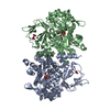
Mass: 62.005 Da / Num. of mol.: 3 / Source method: obtained synthetically / Formula: NO3
#4: Water ChemComp-HOH / water / Water


Mass: 18.015 Da / Num. of mol.: 369 / Source method: isolated from a natural source / Formula: H2O

-
Experimental details

-
Experiment

ExperimentMethod: X-RAY DIFFRACTION / Number of used crystals: 1

-
Sample preparation

CrystalDensity Matthews: 2.24 Å3/Da / Density % sol: 44.89 % / Description: thin plates
Crystal growTemperature: 293 K / Method: vapor diffusion, hanging drop / pH: 6
Details: 34-36% dimethyl PEG 500, 240 mM potassium nitrate, 20 mM magnesium chloride, 100 mM BIS-TRIS, pH 6.0

-
Data collection

DiffractionMean temperature: 100 K
Diffraction sourceSource: SYNCHROTRON / Site: APS / Beamline: 23-ID-B / Wavelength: 0.97933 Å
DetectorType: MARMOSAIC 300 mm CCD / Detector: CCD / Date: Apr 13, 2015
RadiationMonochromator: Double crystal cryo-cooled Si(111) / Protocol: SINGLE WAVELENGTH / Monochromatic (M) / Laue (L): M / Scattering type: x-ray
Radiation wavelengthWavelength: 0.97933 Å / Relative weight: 1
Reflection twin
Crystal-IDIDOperatorDomain-IDFraction
11H, K, L10.878
11-h,-k,l20.122
ReflectionResolution: 2.04→25 Å / Num. obs: 62519 / % possible obs: 99.9 % / Observed criterion σ(F): 0 / Observed criterion σ(I): -3 / Redundancy: 3.4 % / Biso Wilson estimate: 31.3 Å2 / Rmerge(I) obs: 0.115 / Net I/av σ(I): 9.9 / Net I/σ(I): 7.6
Reflection shellResolution: 2.04→2.11 Å / Redundancy: 3.4 % / Rmerge(I) obs: 0.667 / Mean I/σ(I) obs: 2.3 / % possible all: 100

-
Processing

Software
NameVersionClassification
REFMAC5.8.0123refinement
HKL-2000data reduction
HKL-2000data scaling
PHASER2.5.7phasing
RefinementMethod to determine structure: MOLECULAR REPLACEMENT
Starting model: 1ANF
Resolution: 2.042→24.82 Å / Cor.coef. Fo:Fc: 0.946 / Cor.coef. Fo:Fc free: 0.918 / SU B: 6.088 / SU ML: 0.089 / Cross valid method: THROUGHOUT / ESU R: 0.046 / ESU R Free: 0.037 / Details: HYDROGENS HAVE BEEN ADDED IN THE RIDING POSITIONS
RfactorNum. reflection% reflectionSelection details
Rfree0.22084 3134 5 %RANDOM
Rwork0.18036 ---
obs0.18237 59374 95.68 %-
Solvent computationIon probe radii: 0.9 Å / Shrinkage radii: 0.7 Å / VDW probe radii: 1.2 Å
Displacement parametersBiso mean: 28.299 Å2
Baniso -1Baniso -2Baniso -3
1--0.76 Å20 Å2-0.33 Å2
2---13.42 Å2-0 Å2
3---14.18 Å2
Refinement stepCycle: 1 / Resolution: 2.042→24.82 Å
ProteinNucleic acidLigandSolventTotal
Num. atoms7879 0 58 369 8306
Refine LS restraints
Refine-IDTypeDev idealDev ideal targetNumber
X-RAY DIFFRACTIONr_bond_refined_d0.0090.028157
X-RAY DIFFRACTIONr_bond_other_d0.0020.027711
X-RAY DIFFRACTIONr_angle_refined_deg1.3221.97111076
X-RAY DIFFRACTIONr_angle_other_deg1.005317819
X-RAY DIFFRACTIONr_dihedral_angle_1_deg5.83551027
X-RAY DIFFRACTIONr_dihedral_angle_2_deg34.01925.291361
X-RAY DIFFRACTIONr_dihedral_angle_3_deg14.341151365
X-RAY DIFFRACTIONr_dihedral_angle_4_deg14.6171528
X-RAY DIFFRACTIONr_chiral_restr0.0740.21223
X-RAY DIFFRACTIONr_gen_planes_refined0.0050.0219268
X-RAY DIFFRACTIONr_gen_planes_other0.0010.021784
X-RAY DIFFRACTIONr_nbd_refined
X-RAY DIFFRACTIONr_nbd_other
X-RAY DIFFRACTIONr_nbtor_refined
X-RAY DIFFRACTIONr_nbtor_other
X-RAY DIFFRACTIONr_xyhbond_nbd_refined
X-RAY DIFFRACTIONr_xyhbond_nbd_other
X-RAY DIFFRACTIONr_metal_ion_refined
X-RAY DIFFRACTIONr_metal_ion_other
X-RAY DIFFRACTIONr_symmetry_vdw_refined
X-RAY DIFFRACTIONr_symmetry_vdw_other
X-RAY DIFFRACTIONr_symmetry_hbond_refined
X-RAY DIFFRACTIONr_symmetry_hbond_other
X-RAY DIFFRACTIONr_symmetry_metal_ion_refined
X-RAY DIFFRACTIONr_symmetry_metal_ion_other
X-RAY DIFFRACTIONr_mcbond_it1.5172.4554072
X-RAY DIFFRACTIONr_mcbond_other1.5172.4554071
X-RAY DIFFRACTIONr_mcangle_it2.57327.5555090
X-RAY DIFFRACTIONr_mcangle_other2.57327.5575091
X-RAY DIFFRACTIONr_scbond_it2.2062.8364085
X-RAY DIFFRACTIONr_scbond_other2.2062.8314076
X-RAY DIFFRACTIONr_scangle_it
X-RAY DIFFRACTIONr_scangle_other3.79630.515971
X-RAY DIFFRACTIONr_long_range_B_refined5.69361.1579329
X-RAY DIFFRACTIONr_long_range_B_other5.66160.5589230
X-RAY DIFFRACTIONr_rigid_bond_restr
X-RAY DIFFRACTIONr_sphericity_free
X-RAY DIFFRACTIONr_sphericity_bonded
LS refinement shellResolution: 2.042→2.095 Å / Total num. of bins used: 20
RfactorNum. reflection% reflection
Rfree0.26 92 -
Rwork0.216 1941 -
obs--42.52 %
Refinement TLS params.

Method: refined / Refine-ID: X-RAY DIFFRACTION

IDL112)L122)L132)L222)L232)L332)S11 (Å °)S12 (Å °)S13 (Å °)S21 (Å °)S22 (Å °)S23 (Å °)S31 (Å °)S32 (Å °)S33 (Å °)T112)T122)T132)T222)T232)T332)Origin x (Å)Origin y (Å)Origin z (Å)
11.03150.1090.04140.4027-0.36540.45490.0146-0.07680.06630.0371-0.0348-0.0571-0.09180.00270.02020.05210.00040.02470.05340.00260.03459.1542-7.6103-60.7679
20.2719-0.12880.05550.2765-0.30460.5260.02170.0319-0.0534-0.0056-0.03460.0175-0.0322-0.00310.01290.02890.00780.01640.0618-0.01290.0383-0.8348-6.9421-78.431
32.2387-0.28491.29710.44130.32761.3560.01280.0922-0.1545-0.11920.1327-0.0265-0.14770.2045-0.14550.0431-0.020.03410.09510.02560.075723.65532.3325-82.3066
40.9169-0.3044-0.80563.5099-0.69520.994-0.0278-0.30360.04250.406-0.0326-0.2452-0.070.3270.06040.06660.01950.00870.17140.04130.087432.151217.0542-66.5156
50.77980.1913-0.14483.3845-1.58590.773-0.0361-0.44880.17170.6501-0.1472-0.2792-0.24660.14040.18340.24760.08590.05590.3173-0.00720.216931.095214.0893-57.8702
61.27520.1411-0.00370.194-0.36410.7468-0.06380.1252-0.1693-0.02950.0122-0.03930.0618-0.00620.05160.0526-0.02110.03920.0648-0.02410.0427.45763.2802-38.6039
70.21130.13050.04040.2727-0.16360.35950.0127-0.01510.03560.0063-0.01780.00330.0283-0.01370.00510.0298-0.01160.02110.0676-0.00830.0288-2.66112.5923-20.7055
82.73380.0621-0.94350.83230.62130.828-0.08620.09880.1387-0.12180.1627-0.0329-0.05530.0636-0.07640.076-0.02440.01660.10130.04730.051121.8197-7.2329-16.5871
90.88930.82770.95822.67310.51732.5945-0.10210.1151-0.1439-0.1670.0778-0.0895-0.18780.40330.02430.0211-0.03720.0030.13820.01840.048130.1264-22.6267-32.4892
100.9165-1.4342.00473.0795-2.31255.44090.22130.35450.0087-0.757-0.2207-0.2548-0.0581.254-0.00060.341-0.1644-0.01440.36210.03820.274429.8445-12.4696-43.4118
Refinement TLS group
IDRefine-IDRefine TLS-IDAuth asym-IDAuth seq-ID
1X-RAY DIFFRACTION1A-371 - -297
2X-RAY DIFFRACTION2A-296 - -17
3X-RAY DIFFRACTION3A-16 - 514
4X-RAY DIFFRACTION4A515 - 585
5X-RAY DIFFRACTION5A586 - 635
6X-RAY DIFFRACTION6B-371 - -297
7X-RAY DIFFRACTION7B-296 - -18
8X-RAY DIFFRACTION8B-17 - 514
9X-RAY DIFFRACTION9B515 - 600
10X-RAY DIFFRACTION10B601 - 635

+
About Yorodumi

-
News

-
Feb 9, 2022. New format data for meta-information of EMDB entries

New format data for meta-information of EMDB entries

  • Version 3 of the EMDB header file is now the official format.
  • The previous official version 1.9 will be removed from the archive.

Related info.:EMDB header

External links:wwPDB to switch to version 3 of the EMDB data model

-
Aug 12, 2020. Covid-19 info

Covid-19 info

URL: https://pdbj.org/emnavi/covid19.php

New page: Covid-19 featured information page in EM Navigator.

Related info.:Covid-19 info / Mar 5, 2020. Novel coronavirus structure data

+
Mar 5, 2020. Novel coronavirus structure data

Novel coronavirus structure data

Related info.:Yorodumi Speices / Aug 12, 2020. Covid-19 info

External links:COVID-19 featured content - PDBj / Molecule of the Month (242):Coronavirus Proteases

+
Jan 31, 2019. EMDB accession codes are about to change! (news from PDBe EMDB page)

EMDB accession codes are about to change! (news from PDBe EMDB page)

  • The allocation of 4 digits for EMDB accession codes will soon come to an end. Whilst these codes will remain in use, new EMDB accession codes will include an additional digit and will expand incrementally as the available range of codes is exhausted. The current 4-digit format prefixed with “EMD-” (i.e. EMD-XXXX) will advance to a 5-digit format (i.e. EMD-XXXXX), and so on. It is currently estimated that the 4-digit codes will be depleted around Spring 2019, at which point the 5-digit format will come into force.
  • The EM Navigator/Yorodumi systems omit the EMD- prefix.

Related info.:Q: What is EMD? / ID/Accession-code notation in Yorodumi/EM Navigator

External links:EMDB Accession Codes are Changing Soon! / Contact to PDBj

+
Jul 12, 2017. Major update of PDB

Major update of PDB

  • wwPDB released updated PDB data conforming to the new PDBx/mmCIF dictionary.
  • This is a major update changing the version number from 4 to 5, and with Remediation, in which all the entries are updated.
  • In this update, many items about electron microscopy experimental information are reorganized (e.g. em_software).
  • Now, EM Navigator and Yorodumi are based on the updated data.

External links:wwPDB Remediation / Enriched Model Files Conforming to OneDep Data Standards Now Available in the PDB FTP Archive

-
Yorodumi

Thousand views of thousand structures

  • Yorodumi is a browser for structure data from EMDB, PDB, SASBDB, etc.
  • This page is also the successor to EM Navigator detail page, and also detail information page/front-end page for Omokage search.
  • The word "yorodu" (or yorozu) is an old Japanese word meaning "ten thousand". "mi" (miru) is to see.

Related info.:EMDB / PDB / SASBDB / Comparison of 3 databanks / Yorodumi Search / Aug 31, 2016. New EM Navigator & Yorodumi / Yorodumi Papers / Jmol/JSmol / Function and homology information / Changes in new EM Navigator and Yorodumi

Read more