 Movie
Movie Controller


+ Open data

- Basic information
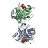
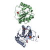
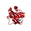
| Entry | Database: PDB / ID: 1idn | ||||||
|---|---|---|---|---|---|---|---|
| Title | MAC-1 I DOMAIN METAL FREE | ||||||
 Components Components | CD11B Integrin alpha M Integrin alpha M | ||||||
 Keywords Keywords | CELL ADHESION / INTEGRIN / I DOMAIN | ||||||
| Function / homology |  Function and homology information Function and homology informationectodermal cell differentiation / integrin alphaM-beta2 complex / positive regulation of prostaglandin-E synthase activity / response to curcumin / positive regulation of neutrophil degranulation / response to Gram-positive bacterium / positive regulation of microglial cell mediated cytotoxicity / complement component C3b binding / vertebrate eye-specific patterning / complement-mediated synapse pruning ...ectodermal cell differentiation / integrin alphaM-beta2 complex / positive regulation of prostaglandin-E synthase activity / response to curcumin / positive regulation of neutrophil degranulation / response to Gram-positive bacterium / positive regulation of microglial cell mediated cytotoxicity / complement component C3b binding / vertebrate eye-specific patterning / complement-mediated synapse pruning / Toll Like Receptor 4 (TLR4) Cascade / negative regulation of dopamine metabolic process / cell-cell adhesion via plasma-membrane adhesion molecules / complement receptor mediated signaling pathway / heterotypic cell-cell adhesion / integrin complex / cargo receptor activity / cell adhesion mediated by integrin / phagocytosis, engulfment / amyloid-beta clearance / plasma membrane raft / tertiary granule membrane / positive regulation of protein targeting to membrane / Integrin cell surface interactions / specific granule membrane / response to mechanical stimulus / forebrain development / heat shock protein binding / receptor-mediated endocytosis / cell-matrix adhesion / positive regulation of superoxide anion generation / response to ischemia / integrin-mediated signaling pathway / Cell surface interactions at the vascular wall / microglial cell activation / cell-cell adhesion / integrin binding / response to estradiol / amyloid-beta binding / Interleukin-4 and Interleukin-13 signaling / cell adhesion / external side of plasma membrane / innate immune response / Neutrophil degranulation / cell surface / extracellular space / extracellular exosome / metal ion binding / plasma membraneSimilarity search - Function | ||||||
| Biological species |  Homo sapiens (human) Homo sapiens (human) | ||||||
| Method | X-RAY DIFFRACTION / MIR / Resolution: 2.7 Å | ||||||
 Authors Authors | Baldwin, E.T. | ||||||
 Citation Citation | Journal: Structure / Year: 1998 Title: Cation binding to the integrin CD11b I domain and activation model assessment Authors: Baldwin, E.T. / Sarver, R.W. / Bryant Jr., G.L. / Curry, K.A. / Fairbanks, M.B. / Finzel, B.C. / Garlick, R.L. / Heinrikson, R.L. / Horton, N.C. / Kelley, L.L. / Mildner, A.M. / Moon, J.B. / ...Authors: Baldwin, E.T. / Sarver, R.W. / Bryant Jr., G.L. / Curry, K.A. / Fairbanks, M.B. / Finzel, B.C. / Garlick, R.L. / Heinrikson, R.L. / Horton, N.C. / Kelley, L.L. / Mildner, A.M. / Moon, J.B. / Mott, J.E. / Mutchler, V.T. / Tomich, C.S. / Watenpaugh, K.D. / Wiley, V.H. | ||||||
| History |
|
- Structure visualization
| Structure viewer | Molecule: MolmilJmol/JSmol |
|---|
- Downloads & links
-Download
| PDBx/mmCIF format | 1idn.cif.gz | 88.4 KB | Display | PDBx/mmCIF format |
|---|---|---|---|---|
| PDB format | pdb1idn.ent.gz | 70.1 KB | Display | PDB format |
| PDBx/mmJSON format | 1idn.json.gz | Tree view | PDBx/mmJSON format | |
| Others |  Other downloads Other downloads |
-Validation report
| Arichive directory | https://data.pdbj.org/pub/pdb/validation_reports/id/1idnftp://data.pdbj.org/pub/pdb/validation_reports/id/1idn | HTTPS FTP |
|---|
-Related structure data


-Links
 PDBj
PDBj


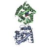
- Assembly
| Deposited unit | 
| ||||||||
|---|---|---|---|---|---|---|---|---|---|
| 1 |
| ||||||||
| Unit cell |
| ||||||||
| Noncrystallographic symmetry (NCS) | NCS oper: (Code: given Matrix: (-0.9633, 0.2683, 0.0089), Vector : |
-Components
| #1: Protein | Integrin alpha M / CELL SURFACE GLYCOPROTEIN MAC-1 ALPHA SUBUNIT Mass: 21689.836 Da / Num. of mol.: 2 / Fragment: MAC-1 ALPHA DOMAIN Source method: isolated from a genetically manipulated source Source: (gene. exp.) Homo sapiens (human) / Cell line: BL21 / Plasmid: BL21 / Production host:  Escherichia coli (E. coli) / Strain (production host): BL21 (DE3) PLYSE / References: UniProt: P11215 Escherichia coli (E. coli) / Strain (production host): BL21 (DE3) PLYSE / References: UniProt: P11215#2: Water | ChemComp-HOH / | Water Mass: 18.015 Da / Num. of mol.: 178 / Source method: isolated from a natural source / Formula: H2O Mass: 18.015 Da / Num. of mol.: 178 / Source method: isolated from a natural source / Formula: H2O |
|---|
-Experimental details
-Experiment
| Experiment | Method: X-RAY DIFFRACTION / Number of used crystals: 1 |
|---|
- Sample preparation
| Crystal | Density Matthews: 2.48 Å3/Da / Density % sol: 50 % Description: DATA WERE COLLECTED BY OSCILLATION WITH 0.25 DEGREE FRAME WIDTHS | ||||||||||||||||||||||||||||||
|---|---|---|---|---|---|---|---|---|---|---|---|---|---|---|---|---|---|---|---|---|---|---|---|---|---|---|---|---|---|---|---|
| Crystal grow | Method: vapor diffusion, sitting drop / pH: 5 Details: CRYSTALS WERE GROWN BY VAPOR DIFFUSION ON SITTING DROP BRIDGES. THE WELL MIX OF 20-24% PEG6000 BUFFERED WITH 100 MM NA ACETATE PH 5.0 WAS MIXED 1:1 WITH 3 UL OF I DOMAIN PROTEIN (20-30 ...Details: CRYSTALS WERE GROWN BY VAPOR DIFFUSION ON SITTING DROP BRIDGES. THE WELL MIX OF 20-24% PEG6000 BUFFERED WITH 100 MM NA ACETATE PH 5.0 WAS MIXED 1:1 WITH 3 UL OF I DOMAIN PROTEIN (20-30 MG/ML, 50 MM HEPES PH 7.0, 0.025% NA AZIDE). CRYSTALS WERE STABLIZED IN 100 MM NA ACETATE 5.0; 26% PEG6000 FOR DATA COLLECTION., vapor diffusion - sitting drop PH range: 5.0-7.0 | ||||||||||||||||||||||||||||||
| Crystal grow | *PLUS Method: vapor diffusion, sitting drop / Details: reservoir was mixed 1:1 with 3 microlitter protein | ||||||||||||||||||||||||||||||
| Components of the solutions | *PLUS
|
-Data collection
| Diffraction | Mean temperature: 287 K |
|---|---|
| Diffraction source | Source: ROTATING ANODE / Type: SIEMENS / Wavelength: 1.5418 |
| Detector | Type: SIEMENS / Detector: AREA DETECTOR / Date: Aug 1, 1994 |
| Radiation | Monochromator: GRAPHITE(002) / Monochromatic (M) / Laue (L): M / Scattering type: x-ray |
| Radiation wavelength | Wavelength: 1.5418 Å / Relative weight: 1 |
| Reflection | Resolution: 2.7→10 Å / Num. obs: 9011 / % possible obs: 68 % / Observed criterion σ(I): 2 / Redundancy: 5.6 % / Rsym value: 0.097 / Net I/σ(I): 16.3 |
| Reflection shell | Resolution: 2.7→2.94 Å / Redundancy: 2.9 % / Mean I/σ(I) obs: 4.6 / Rsym value: 0.173 / % possible all: 39.8 |
| Reflection | *PLUS Rmerge(I) obs: 0.097 |
| Reflection shell | *PLUS % possible obs: 39.8 % / Rmerge(I) obs: 0.173 |
- Processing
| Software |
| ||||||||||||||||||||||||||||||||||||||||||||||||||||||||||||||||||||||||||||||||||||
|---|---|---|---|---|---|---|---|---|---|---|---|---|---|---|---|---|---|---|---|---|---|---|---|---|---|---|---|---|---|---|---|---|---|---|---|---|---|---|---|---|---|---|---|---|---|---|---|---|---|---|---|---|---|---|---|---|---|---|---|---|---|---|---|---|---|---|---|---|---|---|---|---|---|---|---|---|---|---|---|---|---|---|---|---|---|
| Refinement | Method to determine structure: MIR / Resolution: 2.7→10 Å / σ(F): 2 Details: PARAMETERS FROM SIELECKI ET AL. JMB 134, 781-804 1979
| ||||||||||||||||||||||||||||||||||||||||||||||||||||||||||||||||||||||||||||||||||||
| Displacement parameters | Biso mean: 12.49 Å2 | ||||||||||||||||||||||||||||||||||||||||||||||||||||||||||||||||||||||||||||||||||||
| Refinement step | Cycle: LAST / Resolution: 2.7→10 Å
| ||||||||||||||||||||||||||||||||||||||||||||||||||||||||||||||||||||||||||||||||||||
| Refine LS restraints |
| ||||||||||||||||||||||||||||||||||||||||||||||||||||||||||||||||||||||||||||||||||||
| Software | *PLUS Name: PROLSQ / Classification: refinement | ||||||||||||||||||||||||||||||||||||||||||||||||||||||||||||||||||||||||||||||||||||
| Refinement | *PLUS | ||||||||||||||||||||||||||||||||||||||||||||||||||||||||||||||||||||||||||||||||||||
| Solvent computation | *PLUS | ||||||||||||||||||||||||||||||||||||||||||||||||||||||||||||||||||||||||||||||||||||
| Displacement parameters | *PLUS |
