 Movie
Movie Controller

[English] 日本語
 Yorodumi
- PDB-1i3s: THE 2.7 ANGSTROM RESOLUTION CRYSTAL STRUCTURE OF A MUTATED BACULO... -
+ Open data

- Basic information
| Entry | Database: PDB / ID: 1i3s | ||||||
|---|---|---|---|---|---|---|---|
| Title | THE 2.7 ANGSTROM RESOLUTION CRYSTAL STRUCTURE OF A MUTATED BACULOVIRUS P35 AFTER CASPASE CLEAVAGE | ||||||
 Components Components | EARLY 35 KDA PROTEIN | ||||||
 Keywords Keywords | APOPTOSIS / hairpin loop / helix-turn-helix / reactive site loop | ||||||
| Function / homology |  Function and homology information Function and homology informationsymbiont-mediated suppression of host RNAi-mediated antiviral immune response / : / cysteine-type endopeptidase inhibitor activity / cysteine-type endopeptidase inhibitor activity involved in apoptotic process / : / negative regulation of apoptotic process Similarity search - Function | ||||||
| Biological species |  Autographa californica nucleopolyhedrovirus Autographa californica nucleopolyhedrovirus | ||||||
| Method | X-RAY DIFFRACTION / SYNCHROTRON / MOLECULAR REPLACEMENT / Resolution: 2.7 Å | ||||||
 Authors Authors | dela Cruz, W.P. / Lemongello, D. / Friesen, P.D. / Fisher, A.J. | ||||||
 Citation Citation | Journal: J.Biol.Chem. / Year: 2001 Title: Crystal structure of baculovirus P35 reveals a novel conformational change in the reactive site loop after caspase cleavage. Authors: dela Cruz, W.P. / Friesen, P.D. / Fisher, A.J. #1: Journal: Embo J. / Year: 1999Title: Crystal structure of baculovirus P35: role of a novel reactive site loop in apoptotic inhibition Authors: Fisher, A.J. / dela Cruz, W.P. / Zoog, S.J. / Schneider, C.L. / Friesen, P.D. | ||||||
| History |
|
- Structure visualization
| Structure viewer | Molecule: MolmilJmol/JSmol |
|---|
- Downloads & links
-Download
| PDBx/mmCIF format | 1i3s.cif.gz | 177.1 KB | Display | PDBx/mmCIF format |
|---|---|---|---|---|
| PDB format | pdb1i3s.ent.gz | 140.6 KB | Display | PDB format |
| PDBx/mmJSON format | 1i3s.json.gz | Tree view | PDBx/mmJSON format | |
| Others |  Other downloads Other downloads |
-Validation report
| Arichive directory | https://data.pdbj.org/pub/pdb/validation_reports/i3/1i3sftp://data.pdbj.org/pub/pdb/validation_reports/i3/1i3s | HTTPS FTP |
|---|
-Related structure data
| Related structure data |  1i3pSC S: Starting model for refinement C: citing same article ( |
|---|---|
| Similar structure data |


-Links
 PDBj
PDBj


- Assembly
| Deposited unit | 
| ||||||||
|---|---|---|---|---|---|---|---|---|---|
| 1 | 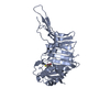
| ||||||||
| 2 | 
| ||||||||
| 3 | 
| ||||||||
| Unit cell |
| ||||||||
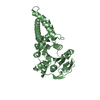
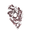
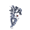
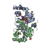
| Details | The biological assembly is a monomer |
-Components
| #1: Protein | / APOPTOSIS PREVENTING PROTEIN Mass: 34743.512 Da / Num. of mol.: 3 / Mutation: YES Source method: isolated from a genetically manipulated source Source: (gene. exp.) Autographa californica nucleopolyhedrovirusGenus: Nucleopolyhedrovirus Alphabaculovirus / Gene: P35 / Plasmid: PET22B+ / Species (production host): Escherichia coli / Production host:  Escherichia coli BL21 (bacteria) / Strain (production host): BL21 / References: UniProt: P08160 Escherichia coli BL21 (bacteria) / Strain (production host): BL21 / References: UniProt: P08160#2: Chemical | Dithiothreitol  Mass: 154.251 Da / Num. of mol.: 3 / Source method: obtained synthetically / Formula: C4H10O2S2 Mass: 154.251 Da / Num. of mol.: 3 / Source method: obtained synthetically / Formula: C4H10O2S2#3: Water | ChemComp-HOH / | Water Mass: 18.015 Da / Num. of mol.: 54 / Source method: isolated from a natural source / Formula: H2O Mass: 18.015 Da / Num. of mol.: 54 / Source method: isolated from a natural source / Formula: H2O |
|---|
-Experimental details
-Experiment
| Experiment | Method: X-RAY DIFFRACTION / Number of used crystals: 1 |
|---|
- Sample preparation
| Crystal | Density Matthews: 2.26 Å3/Da / Density % sol: 45.62 % | ||||||||||||||||||||||||||||||||||||||||||||||||||||||||||||||||||
|---|---|---|---|---|---|---|---|---|---|---|---|---|---|---|---|---|---|---|---|---|---|---|---|---|---|---|---|---|---|---|---|---|---|---|---|---|---|---|---|---|---|---|---|---|---|---|---|---|---|---|---|---|---|---|---|---|---|---|---|---|---|---|---|---|---|---|---|
| Crystal grow | Temperature: 298 K / Method: vapor diffusion, hanging drop / pH: 6 Details: 9-13% PEG 20,000, 100-400 mM NaCl, 100 mM MES, pH 6.0, VAPOR DIFFUSION, HANGING DROP, temperature 298K | ||||||||||||||||||||||||||||||||||||||||||||||||||||||||||||||||||
| Crystal | *PLUS Density % sol: 44.8 % | ||||||||||||||||||||||||||||||||||||||||||||||||||||||||||||||||||
| Crystal grow | *PLUS Temperature: 25 ℃ / pH: 7 | ||||||||||||||||||||||||||||||||||||||||||||||||||||||||||||||||||
| Components of the solutions | *PLUS
|
-Data collection
| Diffraction | Mean temperature: 170 K |
|---|---|
| Diffraction source | Source: SYNCHROTRON / Site: SSRL  / Beamline: BL7-1 / Wavelength: 1.08 Å / Beamline: BL7-1 / Wavelength: 1.08 Å |
| Detector | Type: MARRESEARCH / Detector: IMAGE PLATE / Date: Apr 12, 2000 / Details: mirrors |
| Radiation | Monochromator: Ni MIRROR + Ni FILTER / Protocol: SINGLE WAVELENGTH / Monochromatic (M) / Laue (L): M / Scattering type: x-ray |
| Radiation wavelength | Wavelength: 1.08 Å / Relative weight: 1 |
| Reflection | Resolution: 2.7→30 Å / Num. all: 26627 / Num. obs: 25271 / % possible obs: 94.9 % / Observed criterion σ(F): 0 / Observed criterion σ(I): 0 / Redundancy: 7.26 % / Biso Wilson estimate: 29.3 Å2 / Rmerge(I) obs: 0.07 / Net I/σ(I): 450.7 |
| Reflection shell | Resolution: 2.7→2.8 Å / Redundancy: 3.46 % / Rmerge(I) obs: 0.334 / % possible all: 96.3 |
| Reflection | *PLUS Num. measured all: 183500 / Rmerge(I) obs: 0.07 |
- Processing
| Software |
| ||||||||||||||||||||||||||||||||||||||||||||||||||||||||||||
|---|---|---|---|---|---|---|---|---|---|---|---|---|---|---|---|---|---|---|---|---|---|---|---|---|---|---|---|---|---|---|---|---|---|---|---|---|---|---|---|---|---|---|---|---|---|---|---|---|---|---|---|---|---|---|---|---|---|---|---|---|---|
| Refinement | Method to determine structure: MOLECULAR REPLACEMENT Starting model: PDB ENTRY 1I3P Resolution: 2.7→29.49 Å / Rfactor Rfree error: 0.007 / Data cutoff high absF: 1079019.62 / Data cutoff low absF: 0 / Isotropic thermal model: OVERALL / Cross valid method: THROUGHOUT / σ(F): 0 / σ(I): 0 / Stereochemistry target values: Engh & Huber
| ||||||||||||||||||||||||||||||||||||||||||||||||||||||||||||
| Solvent computation | Solvent model: FLAT MODEL / Bsol: 35.49 Å2 / ksol: 0.322 e/Å3 | ||||||||||||||||||||||||||||||||||||||||||||||||||||||||||||
| Displacement parameters | Biso mean: 35.2 Å2
| ||||||||||||||||||||||||||||||||||||||||||||||||||||||||||||
| Refine analyze |
| ||||||||||||||||||||||||||||||||||||||||||||||||||||||||||||
| Refinement step | Cycle: LAST / Resolution: 2.7→29.49 Å
| ||||||||||||||||||||||||||||||||||||||||||||||||||||||||||||
| Refine LS restraints |
| ||||||||||||||||||||||||||||||||||||||||||||||||||||||||||||
| Refine LS restraints NCS | NCS model details: CONSTR | ||||||||||||||||||||||||||||||||||||||||||||||||||||||||||||
| LS refinement shell | Resolution: 2.7→2.87 Å / Rfactor Rfree error: 0.021 / Total num. of bins used: 6
| ||||||||||||||||||||||||||||||||||||||||||||||||||||||||||||
| Software | *PLUS Name: CNS / Version: 1 / Classification: refinement | ||||||||||||||||||||||||||||||||||||||||||||||||||||||||||||
| Refinement | *PLUS σ(F): 0 / % reflection Rfree: 4.8 % | ||||||||||||||||||||||||||||||||||||||||||||||||||||||||||||
| Solvent computation | *PLUS | ||||||||||||||||||||||||||||||||||||||||||||||||||||||||||||
| Displacement parameters | *PLUS Biso mean: 35.2 Å2 | ||||||||||||||||||||||||||||||||||||||||||||||||||||||||||||
| Refine LS restraints | *PLUS
| ||||||||||||||||||||||||||||||||||||||||||||||||||||||||||||
| LS refinement shell | *PLUS Rfactor Rfree: 0.306 / % reflection Rfree: 4.9 % / Rfactor Rwork: 0.238 |
