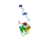
 Movie
Movie Controller


+ Open data

- Basic information
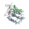
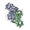
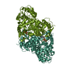
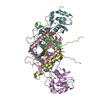
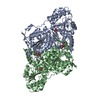
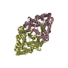
| Entry | Database: PDB / ID: 1i3o | ||||||
|---|---|---|---|---|---|---|---|
| Title | CRYSTAL STRUCTURE OF THE COMPLEX OF XIAP-BIR2 AND CASPASE 3 | ||||||
 Components Components |
| ||||||
 Keywords Keywords |  APOPTOSIS / complex / IAP / caspase APOPTOSIS / complex / IAP / caspase | ||||||
| Function / homology |  Function and homology information Function and homology informationendopeptidase regulator activity / regulation of apoptosis involved in tissue homeostasis / inhibition of cysteine-type endopeptidase activity / positive regulation of protein linear polyubiquitination / regulation of BMP signaling pathway / copper ion homeostasis / nucleotide-binding oligomerization domain containing 1 signaling pathway / regulation of nucleotide-binding domain, leucine rich repeat containing receptor signaling pathway / caspase-3 / Stimulation of the cell death response by PAK-2p34 ...endopeptidase regulator activity / regulation of apoptosis involved in tissue homeostasis / inhibition of cysteine-type endopeptidase activity / positive regulation of protein linear polyubiquitination / regulation of BMP signaling pathway / copper ion homeostasis / nucleotide-binding oligomerization domain containing 1 signaling pathway / regulation of nucleotide-binding domain, leucine rich repeat containing receptor signaling pathway / caspase-3 / Stimulation of the cell death response by PAK-2p34 / phospholipase A2 activator activity / anterior neural tube closure / intrinsic apoptotic signaling pathway in response to osmotic stress / leukocyte apoptotic process / positive regulation of pyroptotic inflammatory response / glial cell apoptotic process / NADE modulates death signalling / luteolysis / response to cobalt ion / cysteine-type endopeptidase activity involved in apoptotic signaling pathway / protein serine/threonine kinase binding / death-inducing signaling complex / cyclin-dependent protein serine/threonine kinase inhibitor activity / cellular response to staurosporine / Apoptosis induced DNA fragmentation / nucleotide-binding oligomerization domain containing 2 signaling pathway / cysteine-type endopeptidase activity involved in execution phase of apoptosis / Caspase activation via Dependence Receptors in the absence of ligand / Apoptotic cleavage of cell adhesion proteins / death receptor binding / SMAC, XIAP-regulated apoptotic response / axonal fasciculation / Regulation of the apoptosome activity / Activation of caspases through apoptosome-mediated cleavage / Signaling by Hippo / SMAC (DIABLO) binds to IAPs / SMAC(DIABLO)-mediated dissociation of IAP:caspase complexes / cysteine-type endopeptidase activity involved in apoptotic process / TNFR1-induced proapoptotic signaling / fibroblast apoptotic process / execution phase of apoptosis / negative regulation of cytokine production / epithelial cell apoptotic process / platelet formation / RIPK1-mediated regulated necrosis / cysteine-type endopeptidase inhibitor activity / Other interleukin signaling / positive regulation of amyloid-beta formation / regulation of innate immune response / pyroptotic inflammatory response / Apoptotic cleavage of cellular proteins / negative regulation of B cell proliferation / T cell homeostasis / negative regulation of activated T cell proliferation / protein K63-linked ubiquitination / cysteine-type endopeptidase inhibitor activity involved in apoptotic process / neurotrophin TRK receptor signaling pathway / B cell homeostasis / protein maturation / negative regulation of cell cycle / response to X-ray / positive regulation of type I interferon production / negative regulation of tumor necrosis factor-mediated signaling pathway / Caspase-mediated cleavage of cytoskeletal proteins / regulation of macroautophagy / response to amino acid / cell fate commitment / Pyroptosis / response to tumor necrosis factor / response to glucose / response to UV / response to glucocorticoid / keratinocyte differentiation / striated muscle cell differentiation / Degradation of the extracellular matrix / Regulation of PTEN localization / intrinsic apoptotic signaling pathway / erythrocyte differentiation / response to nicotine / TNFR1-induced NF-kappa-B signaling pathway / positive regulation of protein ubiquitination / Deactivation of the beta-catenin transactivating complex / apoptotic signaling pathway / hippocampus development / Regulation of TNFR1 signaling / sensory perception of sound / positive regulation of JNK cascade / protein catabolic process / regulation of protein stability / response to hydrogen peroxide / RING-type E3 ubiquitin transferase / negative regulation of cysteine-type endopeptidase activity involved in apoptotic process / Regulation of necroptotic cell death / protein processing / neuron differentiation / Wnt signaling pathway / response to wounding / Regulation of PTEN stability and activity / ubiquitin-protein transferase activity / positive regulation of canonical Wnt signaling pathwaySimilarity search - Function | ||||||
| Biological species |  Homo sapiens (human) Homo sapiens (human) | ||||||
| Method | X-RAY DIFFRACTION / SYNCHROTRON / Resolution: 2.7 Å | ||||||
 Authors Authors | Riedl, S.J. / Renatus, M. / Schwarzenbacher, R. / Zhou, Q. / Sun, C. / Fesik, S.W. / Liddington, R.C. / Salvesen, G.S. | ||||||
 Citation Citation | Journal: Cell(Cambridge,Mass.) / Year: 2001 Title: Structural basis for the inhibition of caspase-3 by XIAP. Authors: Riedl, S.J. / Renatus, M. / Schwarzenbacher, R. / Zhou, Q. / Sun, C. / Fesik, S.W. / Liddington, R.C. / Salvesen, G.S. | ||||||
| History |
|
- Structure visualization
| Structure viewer | Molecule: MolmilJmol/JSmol |
|---|
- Downloads & links
-Download
| PDBx/mmCIF format | 1i3o.cif.gz | 150.7 KB | Display | PDBx/mmCIF format |
|---|---|---|---|---|
| PDB format | pdb1i3o.ent.gz | 117.8 KB | Display | PDB format |
| PDBx/mmJSON format | 1i3o.json.gz | Tree view | PDBx/mmJSON format | |
| Others |  Other downloads Other downloads |
-Validation report
| Arichive directory | https://data.pdbj.org/pub/pdb/validation_reports/i3/1i3oftp://data.pdbj.org/pub/pdb/validation_reports/i3/1i3o | HTTPS FTP |
|---|
-Related structure data


-Links
 PDBj
PDBj


- Assembly
| Deposited unit | 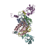
| ||||||||
|---|---|---|---|---|---|---|---|---|---|
| 1 |
| ||||||||
| Unit cell |
|
-Components
| #1: Protein | Mass: 19727.279 Da / Num. of mol.: 2 / Fragment: APOPAIN P17 SUBUNIT / Mutation: C285A Source method: isolated from a genetically manipulated source Source: (gene. exp.) Homo sapiens (human) / Gene: I39005 / Plasmid: PET23B / Species (production host): Escherichia coli / Production host:  Escherichia coli BL21(DE3) (bacteria) / Strain (production host): BL21(DE3) Escherichia coli BL21(DE3) (bacteria) / Strain (production host): BL21(DE3)References: UniProt: P42574, Hydrolases; Acting on peptide bonds (peptidases); Cysteine endopeptidases#2: Protein | Mass: 12981.756 Da / Num. of mol.: 2 / Fragment: APOPAIN P12 SUBUNIT Source method: isolated from a genetically manipulated source Source: (gene. exp.) Homo sapiens (human) / Gene: I39005 / Plasmid: PET23B / Species (production host): Escherichia coli / Production host: Escherichia coli BL21(DE3) (bacteria) / Strain (production host): BL21(DE3)References: UniProt: P42574, Hydrolases; Acting on peptide bonds (peptidases); Cysteine endopeptidases#3: Protein | Mass: 13993.578 Da / Num. of mol.: 2 / Fragment: XIAP-BIR2 / Mutation: C202A, C213G Source method: isolated from a genetically manipulated source Source: (gene. exp.) Homo sapiens (human) / Gene: U32974 / Plasmid: PET15B / Species (production host): Escherichia coli / Production host: Escherichia coli BL21(DE3) (bacteria) / Strain (production host): BL21(DE3) / References: UniProt: P98170#4: Chemical |   Mass: 65.409 Da / Num. of mol.: 2 / Source method: obtained synthetically / Formula: Zn Mass: 65.409 Da / Num. of mol.: 2 / Source method: obtained synthetically / Formula: Zn#5: Water | ChemComp-HOH / | Water Mass: 18.015 Da / Num. of mol.: 13 / Source method: isolated from a natural source / Formula: H2O Mass: 18.015 Da / Num. of mol.: 13 / Source method: isolated from a natural source / Formula: H2O |
|---|
-Experimental details
-Experiment
| Experiment | Method: X-RAY DIFFRACTION / Number of used crystals: 1 |
|---|
- Sample preparation
| Crystal | Density Matthews: 2.61 Å3/Da / Density % sol: 60.14 % | ||||||||||||||||||||||||||||||||||||||||||
|---|---|---|---|---|---|---|---|---|---|---|---|---|---|---|---|---|---|---|---|---|---|---|---|---|---|---|---|---|---|---|---|---|---|---|---|---|---|---|---|---|---|---|---|
| Crystal grow | Temperature: 295 K / Method: vapor diffusion, sitting drop / pH: 5.2 Details: sodium formate, sodium acetate, pH 5.2, VAPOR DIFFUSION, SITTING DROP, temperature 295K | ||||||||||||||||||||||||||||||||||||||||||
| Crystal grow | *PLUS Temperature: 23 ℃ / pH: 7.6 | ||||||||||||||||||||||||||||||||||||||||||
| Components of the solutions | *PLUS
|
-Data collection
| Diffraction | Mean temperature: 100 K |
|---|---|
| Diffraction source | Source: SYNCHROTRON / Site: SSRL  / Beamline: BL7-1 / Wavelength: 1.08 Å / Beamline: BL7-1 / Wavelength: 1.08 Å |
| Detector | Type: MARRESEARCH / Detector: IMAGE PLATE / Date: Dec 15, 2000 |
| Radiation | Protocol: SINGLE WAVELENGTH / Monochromatic (M) / Laue (L): M / Scattering type: x-ray |
| Radiation wavelength | Wavelength: 1.08 Å / Relative weight: 1 |
| Reflection | Resolution: 2.7→500 Å / Num. all: 174633 / Num. obs: 27119 / % possible obs: 99.4 % / Redundancy: 5 % / Biso Wilson estimate: 30 Å2 / Rmerge(I) obs: 0.062 |
| Reflection shell | Resolution: 2.7→2.8 Å / Redundancy: 4.5 % / Rmerge(I) obs: 0.424 / Mean I/σ(I) obs: 2.3 / Num. unique all: 2649 / % possible all: 99.3 |
| Reflection | *PLUS Lowest resolution: 500 Å / Num. measured all: 174633 |
| Reflection shell | *PLUS % possible obs: 99.3 % |
- Processing
| Software |
| ||||||||||||||||||||||||||||||||||||||||||||||||||||||||||||
|---|---|---|---|---|---|---|---|---|---|---|---|---|---|---|---|---|---|---|---|---|---|---|---|---|---|---|---|---|---|---|---|---|---|---|---|---|---|---|---|---|---|---|---|---|---|---|---|---|---|---|---|---|---|---|---|---|---|---|---|---|---|
| Refinement | Starting model: PDB ENTRY 1PAU and PDB ENTRY 1C9Q Resolution: 2.7→500 Å / Cross valid method: THROUGHOUT / σ(F): 0 / σ(I): 0 / Stereochemistry target values: Engh & Huber / Details: MLF
| ||||||||||||||||||||||||||||||||||||||||||||||||||||||||||||
| Solvent computation | Solvent model: CNS BULK SOLVENT / Bsol: 33.0707 Å2 / ksol: 0.321299 e/Å3 | ||||||||||||||||||||||||||||||||||||||||||||||||||||||||||||
| Displacement parameters | Biso mean: 29.938 Å2
| ||||||||||||||||||||||||||||||||||||||||||||||||||||||||||||
| Refinement step | Cycle: LAST / Resolution: 2.7→500 Å
| ||||||||||||||||||||||||||||||||||||||||||||||||||||||||||||
| Refine LS restraints |
| ||||||||||||||||||||||||||||||||||||||||||||||||||||||||||||
| Xplor file |
| ||||||||||||||||||||||||||||||||||||||||||||||||||||||||||||
| Software | *PLUS Name: CNS / Version: 1 / Classification: refinement | ||||||||||||||||||||||||||||||||||||||||||||||||||||||||||||
| Refinement | *PLUS Highest resolution: 2.7 Å / Lowest resolution: 500 Å / σ(F): 0 / % reflection Rfree: 5 % / Rfactor obs: 0.248 | ||||||||||||||||||||||||||||||||||||||||||||||||||||||||||||
| Solvent computation | *PLUS | ||||||||||||||||||||||||||||||||||||||||||||||||||||||||||||
| Displacement parameters | *PLUS | ||||||||||||||||||||||||||||||||||||||||||||||||||||||||||||
| Refine LS restraints | *PLUS
|
